Article Text

Abstract
Purpose The association of human leucocyte antigen (HLA) class I expression levels with the clinical course of many malignancies reflects their crucial role in the recognition and elimination of malignant cells by cognate T cells and NK cells. In colorectal cancer, results regarding this association are conflicting. The potential pathogenetic and therapeutic implications of this association prompted us to perform a large patient-level pooled analysis assessing the role of the expression level of HLA class I loci gene products in colon and rectal cancer.
Experimental design Included studies provided patient-level data on HLA class I expression levels determined by immunohistochemistry on surgical specimens. Expression levels of the HLA class I loci gene products (HLA-A, HLA-B/C) were correlated with common genetic events and survival.
Results Data from 5 studies including 2863 patients were used. In the 1620 colon cancer patients, lower HLA-A, HLA-B/C and total HLA class I expression levels were associated with microsatellite instability (p=0.044, p=0.008 and p=0.022, respectively), higher frequency of BRAF mutations (p<0.001, p=0.021 and p<0.001, respectively) and lower frequency of KRAS mutations (p=0.001, ns and p=0.002, respectively). In the 1243 rectal cancer patients, HLA-A expression was higher in tumors treated with neoadjuvant radiation (p=0.024). High HLA-B/C, but not HLA-A, expression level was an independent predictor of favorable overall survival in colon (p=0.006) and rectal (p<0.001) cancer.
Conclusions T-cells and HLA-B/C antigens, rather than NK cells and HLA-A antigens, likely play an important role in controlling colon/rectal cancer growth. Colon/rectal cancer patients may benefit from strategies that upregulate HLA-B/C and trigger or enhance T cell immunity.
- gastrointestinal neoplasms
- receptors, immunologic
- T-lymphocytes
- adaptive immunity
Data availability statement
All data relevant to the study are included in the article or uploaded as online supplemental information.
This is an open access article distributed in accordance with the Creative Commons Attribution Non Commercial (CC BY-NC 4.0) license, which permits others to distribute, remix, adapt, build upon this work non-commercially, and license their derivative works on different terms, provided the original work is properly cited, appropriate credit is given, any changes made indicated, and the use is non-commercial. See http://creativecommons.org/licenses/by-nc/4.0/.
Statistics from Altmetric.com
Introduction
The impressive clinical responses to checkpoint inhibitor-based immunotherapy in a subset of cancer patients has restored tumor immunologists’ confidence in the role of immunosurveillance in the pathogenesis and clinical course of cancer.1 2 The renewed appreciation of the immune system’s role in oncology has in turn stimulated interest in the characterization of the expression of molecules by cancer cells which mediate their interactions with a host’s immune system. Among them are human leucocyte antigen (HLA) class I molecules which mediate the recognition and elimination of malignant cells by cognate T cells, by presenting tumor antigen (TA)-derived peptides.3 HLA class I loss or downregulation results in impaired recognition and destruction of cancer cells by cognate T cells, thus providing them with an immuno-escape mechanism.4 The clinical relevance of these findings is indicated by the association of HLA class I expression levels on malignant cells with the clinical outcomes of many types of cancer.5
In colorectal cancer (CRC), at least 24 single-institution studies have described defective HLA class I expression with conflicting results regarding the association of HLA class I expression levels with prognosis.6 In some studies, high HLA class I expression levels are associated with a favorable prognosis, implying a role of TA-specific T cell immune responses in the clinical course of the disease.7 8 In other studies, complete HLA class I loss by cancer cells is associated with a favorable prognosis, implying cancer cell elimination by NK cells.9 This bimodal association of HLA class I expression levels with prognosis is not unique of CRC, since it has been described also in breast, cervical and penile cancers.10–13
Characterization of the role of HLA class I expression levels in the interaction of CRC cells with the host’s immune system may assist in prognostication and in selection of the most appropriate immunotherapeutic strategy. Therefore, taking advantage of patient-level data provided by 5 independent studies7–9 14 15 comprizing approximately 3000 patients, we investigated the association of HLA-A and HLA-B,C expression levels—not detected, low, high—with various clinicopathologic variables and survival outcomes. We investigated the effect of total HLA class I expression level and HLA class I loci gene products (HLA-A and HLA-B/C), as they are regulated by different mechanisms and display different functional properties.16 17 To further increase the discriminatory power of our study, we analyzed colon and rectal cancers as distinct entities, due to their unique tumor biologies and clinicopathologic features.18 Lastly, we determined the relationship between HLA class I expression levels and common genetic events, namely microsatellite instability (MSI), KRAS mutations and BRAF mutations,19 as well as neoadjuvant radiation therapy, in order to determine their potential role in HLA class I regulation.
Methods
Study selection
PubMed and Google Scholar databases were searched for studies investigating the association of HLA class I expression level with survival in CRC patients. The following search terms were used: ((((((“hla”) OR “hla a”) OR “hla b”) OR “hla c”) OR “hla class i”)) AND ((“Colorectal Neoplasms”(MeSH)) OR (((((((“colon”) OR “colonic”) OR “rectum”) OR “rectal”) OR “colorectal”)) AND ((((“cancer”) OR “tumor”) OR “malignancy”) OR “neoplasm”))). Studies were considered eligible if they provided information on both HLA class I expression level—including ‘lack’ of expression—in surgically resected colon/rectal cancer specimens and on patient postoperative survival. Studies which did not provide data on ‘lack’ of HLA class I expression were excluded. The literature was searched independently by two investigators (TM and LC). In case of discrepancy, the studies were re-evaluated by a third investigator (FK) until consensus was reached. For each eligible study, the corresponding author was contacted and patient-level data on clinicopathological characteristics, HLA class I expression level, neoadjuvant radiation therapy, MSI, KRAS and BRAF mutations, as well as survival were requested. Obtained individual patient-level data from all studies were combined into a single dataset. Given the different methods used by each individual study to score overall HLA class I expression level, we combined HLA-A, HLA-B/C expression and immunoreactivity score (IRS) in order to categorize total HLA class I expression into ‘loss’, ‘downregulation’ and ‘expression’ (online supplemental table 1).
Supplemental material
Statistical analysis
Continuous variables are presented as median (IQR) and categorical as N (%). Comparison of categorical variables among groups was performed using Fisher’s exact test. Sensitivity analyses were carried out using the leave-one-out approach. Follow-up duration was calculated with the reverse Kaplan-Meier method. disease-free survival (DFS), overall survival (OS) and disease-specific survival (DSS) were defined as the interval between the date of operation and the date of recurrence, any death, or disease-specific death, respectively (event), or the date of last follow-up (censored). Survival curves were plotted using the Kaplan-Meier method. Differences in DFS, OS, and DSS among groups were analyzed by the log-rank test. Multivariable survival analyses were performed using a stepwise backward Cox proportional hazards survival model. The assumption of proportionality for variables included in the Cox analysis was verified by graphical assessment of Kaplan-Meier curves. A p<0.05 was considered statistically significant. All tests were two-tailed. Statistical analyses were performed with StataSE V.15 (StataCorp) and GraphPad Prism, V.8.0 for Windows (GraphPad Software, La Jolla, California, USA).
Results
Characteristics of included studies and patients
We identified 1013 studies in the literature providing information on both HLA class I expression levels—including ‘lack’ of expression—and survival in surgically resected patients with CRC The majority of these studies were excluded because they did not provide information on both CRC and HLA class I expression level (n=989), on survival (n=14), or on ‘lack’ of HLA class I expression (n=5). Ultimately, our analysis included five observational studies for which patient-level HLA class I expression and survival data were available (figure 1, online supplemental table 2). Three studies analyzed HLA-A expression levels using the monoclonal antibody (mAb) HC-A2, while four studies analyzed HLA-B/C expression levels using the mAb HC-10. One study evaluated HLA class I expression levels using the commercially available mouse IgG mAb (MBL, Nagoya, Japan). To quantitate expression levels, four studies measured the percentage of stained cancer cells, while one study analyzed total HLA class I expression levels using the IRS system. The latter takes into account the percentage of stained cancer cells and the staining intensity (percentage (range 0–4) × intensity (range 0–3)).20 Two studies provided microsatellite status data in colon cancer; one provided BRAF status information in colon cancer; two studies in colon cancer and two in rectal cancer provided KRAS data. DFS, OS and DSS were provided to varying degrees.
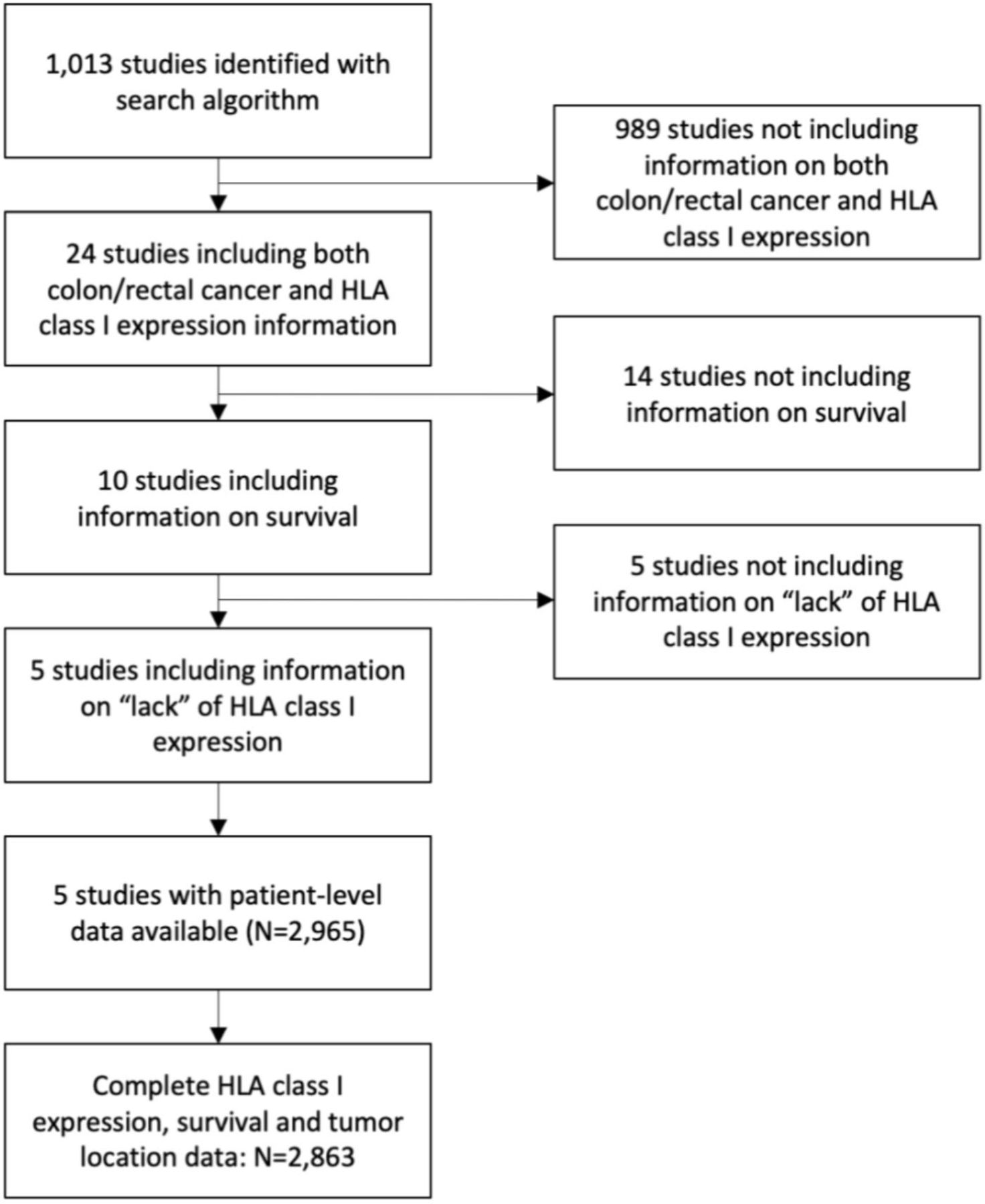
Study selection algorithm. HLA, human leucocyte antigen.
Of the 2965 included patients, 2863 had complete data on tumor location (colon/rectum), HLA class I expression level, and survival. Tumors were located in the colon and rectum in 1620 (57%) and in 1243 (43%) patients, respectively. Patient characteristics by tumor location are summarized in online supplemental table 3.
HLA class I expression level
In colon cancer, HLA-A, HLA-B/C and total HLA class I expression was not detected in 184 (16%), 87 (6%) and 338 (21%) tumors, respectively. In rectal cancer, HLA-A, HLA-B/C and total HLA class I expression was not detected in 80 (8%), 39 (3%) and 120 (10%) tumors, respectively (table 1).
HLA class I expression level in colon or rectal malignant tumors surgically resected from 2863 patients, by tumor location
HLA-A and HLA-B/C expression levels were positively correlated with each other in both colon (p<0.001) and rectal (p<0.001) cancer. HLA-B/C expression levels were significantly higher than that of HLA-A in both colon (p<0.001, figure 2A) and rectal (p<0.001, figure 2B) cancers. HLA-A, HLA-B/C and total HLA class I expression levels were higher in rectal than in colon cancer (p<0.001 for all comparisons, figure 2C–E).
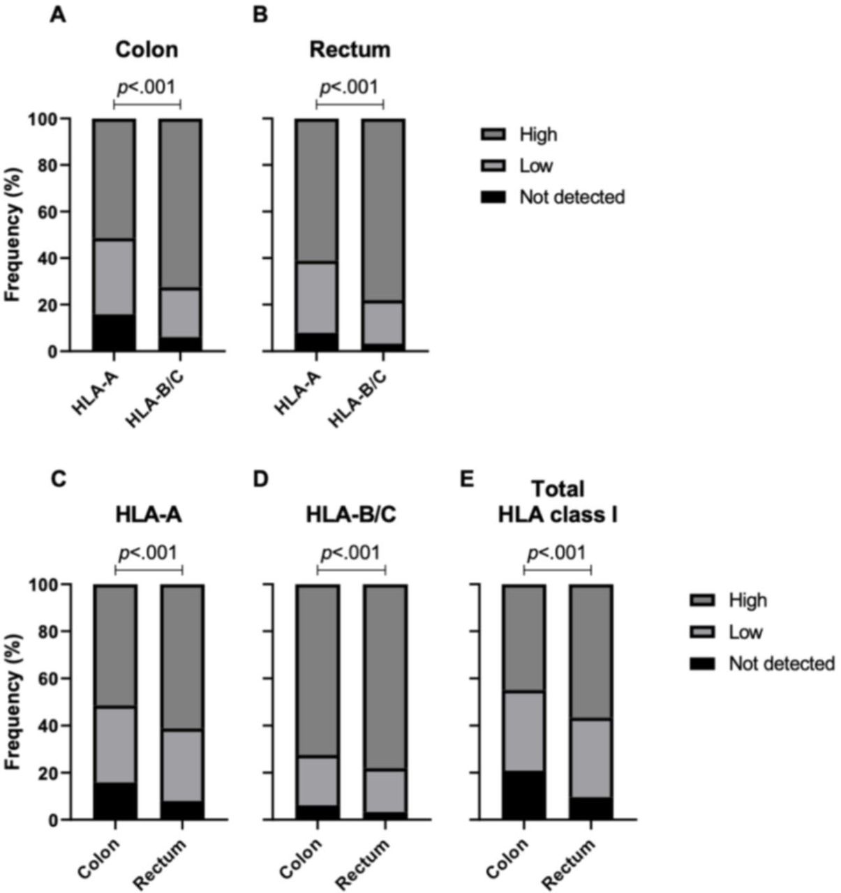
HLA class I expression by qualitative level in colon and rectal malignant tumors. Stacked bar graphs demonstrate higher expression level of HLA-A vs HLA-B/C in colon (A) and rectal (B) malignant tumors, as well as higher HLA-A (C), HLA-B/C (D) and total HLA class I (E) expression level in rectal versus colon malignant tumors. P values were derived from Fisher’s exact test. Qualitative levels included ‘not detected’, ‘low’ and ‘high’ HLA class I, HLA-A and HLA-B/C expression. HAL, human leucocyte antigen.
Association of HLA class I expression levels with clinicopathologic characteristics, mutations and neoadjuvant radiation
In colon cancer, higher HLA-A (p<0.001, online supplemental figure 1A) and total HLA class I (p<0.001, online supplemental figure 1B) expression levels were associated with lower histologic grade, while higher HLA-B/C expression levels were associated with lower stage (p=0.019, online supplemental figure 1C). No associations were observed in rectal cancer.
Lower HLA-A (p=0.044, figure 3A), HLA-B/C (p=0.008, figure 3B), and total HLA class I (p=0.022, figure 3C) expression levels were associated with MSI in colon cancer. Moreover, lower HLA-A (p<0.001, figure 3D), HLA-B/C (p=0.021, figure 3E) and total HLA class I (p<0.001, figure 3F) expression levels were associated with BRAF mutations in colon cancer. In contrast, higher HLA-A (p=0.001, figure 3G) and total HLA class I (p=0.002, figure 3H) expression levels were associated with presence of KRAS mutations in colon cancer.

Association of HLA class I expression level with microsatellite stability (MSS) status, BRAF mutations and KRAS mutations in colon malignant tumors. Stacked bar graphs demonstrate the association of lower HLA-A (A, D), HLA-B/C (B, E), and total HLA class I (C, F) expression levels with microsatellite instability (MSI) and BRAF mutations, and the association of higher HLA-A (G) and total HLA class I (H) expression levels with KRAS mutations in colon malignant tumors. P values were derived from Fisher’s exact test. HLA, human leucocyte antigen;wt, wild type; mut, mutated.
In rectal cancer, HLA-A expression was higher in patients who had received neoadjuvant radiation therapy (p=0.024).
All of the above correlations remained significant when the study by Kasajima et al was excluded from the analysis because of the use of a different antibody and of a different scoring method (data not shown).
Survival
Median follow-up was 53 months (IQR 19–84) for colon cancer and 65 months (IQR 32–85) for rectal cancer patients. Median DFS, OS, and DSS were 78 months (IQR 19-not reached (NR)), 80 months (IQR 23–NR) and NR (IQR 26–NR) for colon cancer, and 113 months (IQR 30–NR), 92 months (IQR 34–NR) and NR (IQR 43–NR) for rectal cancer, respectively.
In colon cancer, higher HLA-B/C expression levels were associated with longer DFS (high: median DFS 86 months (IQR 22–NR) vs low: median DFS 64 months (IQR 14–NR) vs not detected: median DFS 39 months (IQR 13-NR); p=0.014, figure 4B) and OS (high: median OS 90 m (IQR 27–NR) vs low: median OS 64 months (IQR 14–NR) vs not detected: median OS 43 months (IQR 14–118); p<0.001, figure 4E). In contrast, HLA-A and total HLA-class I expression levels were associated neither with DFS (p=0.764, figure 4A, and p=0.279, figure 4C, respectively) nor with OS (p=0.763, figure 4D, and p=0.442, figure 4F, respectively). Neither HLA-A (p=0.804), HLA-B/C (p=0.235), nor total HLA class I (p=0.338) expression levels were associated with DSS.

Association of HLA class I expression level in colon malignant tumors with survival. Kaplan-Meier curves demonstrate the association of HLA-A (A, D), HLA-B/C (B, E) and total HLA class I (C, F) expression levels in colon malignant tumors with overall (A, B, C) and disease-free survival (D, E, F). P values were derived from log-rank tests. HLA, human leucocyte antigen; DFS, disease-free survival; OS, overall survival.
In rectal cancer, higher HLA-B/C expression levels were associated with longer OS (high: median OS 95 months (IQR 36-NR) vs negative: median OS 37 months (IQR 8–59); p<0.001, figure 5B) and DSS (high: median DSS NR (IQR 46–NR) vs low: median DSS 48 months, IQR 17–NR; p<0.001, figure 5E). HLA-A and total HLA class I expression levels were associated neither with OS (p=0.763, p=0.969, respectively, figure 5A,C) nor with DSS (p=0.845, p=0.517, respectively, figure 5D,F). DFS data were not available for rectal cancer. The above results remained unchanged when the study by Kasajima et al was excluded from the analysis because of the use of a different antibody and a different scoring method (data not shown). Presence of KRAS mutations was also associated with shorter OS (KRASwt: median OS NR (IQR 68–NR) vs KRASmut: median OS 56 months (IQR 25–NR); p=0.015).

Association of HLA class I expression level in rectal malignant tumors with survival. Kaplan-Meier curves demonstrate the association of HLA-A (A, D), HLA-B/C (B, E) and total HLA class I (C, F) expression levels in rectal malignant tumors with overall (A, B, C) and disease-specific survival (D, E, F). P values were derived from log-rank tests. HLA, human leucocyte antigen; DFS, disease-free survival; OS, overall survival.
On multivariable analyses for colon cancer patients, lower HLA-B/C expression levels were an independent predictor of shorter DFS (HR=0.8, p=0.006) and OS (HR=0.8, p<0.001) after adjusting for clinicopathologic parameters (table 2). In contrast, HLA-A and total HLA class I expression levels were associated neither with DFS (p=0.940 and p=0.492, respectively) nor with OS (p=0.554 and p=0.747, respectively). Neither HLA-A (p=0.802), HLA-B/C (p=0.627), nor total HLA class I (p=0.748) expression levels were associated with DSS.
Variables associated with survival on univariable and multivariable analyses in 1620 patients with colon and 1243 patients with rectal cancer, by tumor location
In rectal tumors, lower HLA-B/C expression levels emerged as an independent predictor of shorter OS (HR=0.8, p<0.001) after adjusting for clinicopathological factors (table 2). No changes were observed in multivariable analysis results when the study by Kasajima et al was excluded from the analysis because of the use of a different antibody and a different scoring method (data not shown).
Discussion
To assess in a conclusive way the role of HLA class I expression levels in the pathogenesis and clinical course of colon and rectal cancer, we performed a large multi-institutional patient-level pooled analysis including published and novel data from five studies. This analysis which has used data derived from the characterization of approximately 3000 patients with colon or rectal cancer has generated the following conclusions: first, the expression levels of the gene products of HLA class I loci are differentially associated with survival in patients with colon or rectal cancer. Second, lack of HLA class I expression by malignant cells is not associated with a survival benefit. Lastly, HLA class I expression levels are associated with several common genetic events in colon cancer, while in rectal cancer HLA class I expression levels are higher on cancer cells in patients who have received neoadjuvant radiation therapy.
The information in the literature regarding the association between lack of HLA class I expression and survival in colon and other cancers is conflicting. This association has been interpreted to reflect the major role played by NK cells in the clinical course of the disease, since the lack of interactions between HLA class I alleles expressed on malignant cells and killer-cell immunoglobulin-like receptors expressed on NK cells, would not inhibit their ability to eliminate malignant cells.21 Our analysis of 2863 patients demonstrated that lack of HLA-B/C expression was associated with the shortest survival. This association argues against NK cells playing a major role in controlling the growth of malignant cells in colon cancer9–13 but rather implies that T cells play a major role in the clinical course of colon cancer. If the described association is not a fortuitous event and our interpretation is correct, colon cancer patients might benefit from strategies which enhance the T cell immune response to malignant cells.
We also demonstrated a differential association of HLA class I gene products with survival: high HLA-B/C was found to be an independent predictor of favorable prognosis for both colon and rectal cancer, while HLA-A or total HLA class I expression levels were not associated with the clinical course of these diseases. If this association reflects a causal relationship, this finding supports the possibility that HLA-B/C antigens, but not HLA-A antigens, present relevant neoantigens to T cells and mediate an antitumor immune response. This notion is also supported by the fact that HLA-B molecules possibly present a more diverse peptide repertoire compared with HLA-A.17 Thus, HLA-A loss or downregulation is not expected to negatively impact prognosis in colon/rectal cancer. On the other hand, HLA-B/C will need to be evaluated for prognostication purposes. If our interpretation is correct, the differential role of the gene products of HLA class I loci in the clinical course of colon cancer emphasizes the need to assess the expression level of HLA class I loci gene products separately using distinct antibodies with the appropriate specificity. This approach is also supported by the finding that antibodies recognizing total HLA class I expression do not detect selective changes in the expression of HLA class I loci products or alleles.22 23
Additionally, we found that BRAF mutations are associated with lower HLA-A, HLA-B/C, and HLA class I expression levels on malignant colon cancer cells. This association may be mediated by the inhibition of HLA class I expression on colon cancer cells by the MAPK pathway.24 The underlying mechanism is represented by the lack of activation of STAT1 and STAT3, which regulate the transcription of HLA class I antigen processing machinery (APM) components.25 26 An alternative, but not exclusive mechanism, is represented by the rapid internalization and intracellular sequestration of HLA class I on tumor cells induced by BRAF.27 In contrast KRAS mutations, which act upstream of BRAF in the MAPK signaling cascade, were associated with higher HLA-A and total HLA class I expression levels. This association may reflect the involvement of KRAS in the activation of NF-κB signaling in CRC cells.28 29 The latter enhances HLA-A transcription to a greater extent than HLA-B and HLA-C transcription; this differential effect provides a potential mechanism for the selective association of KRAS mutations with higher HLA-A, but not HLA-B/C expression level.30 Lastly, MSI was associated with lower HLA-A, HLA-B/C, and total HLA class I expression levels. This association may reflect a greater frequency of HLA class I heavy chains, β2-microglobulin, transporter associated with antigen processing, and NLRC5 mutations in colorectal tumors displaying MSI.31
Interestingly, we found higher HLA-A expression levels on rectal tumors that had been subjected to neoadjuvant radiation therapy. Although HLA class I upregulation by radiation has been rarely reported in the clinical setting, several in vitro and mouse studies have demonstrated HLA class I upregulation following irradiation.32 33 This effect may be mediated by induction of a type I interferon (IFN) response and enhanced IFN-γ secretion by lymphocytes.32 34 In the setting of irradiation, cGAS/STING and RIG-I/MAVS activation33 35 may contribute to a type I IFN transcription via activation of IRF3, IRF7, and NF-κB. Moreover, ATM kinase and non-canonical STING activation36 37 may activate STAT1 and NF-κB, respectively, driving IFN-independent HLA class I APM component transcription.
Our analysis is limited by the lack of standardization of the methods used and the heterogeneity of patient populations enrolled in the included studies. However, four of the five included studies used the same mAb for the IHC analysis of HLA-A and HLA-B/C expression by malignant cells. Furthermore, we controlled for the individual studies in multivariable analyses, and we performed sensitivity analyses excluding the study which differed the most from the remaining ones in terms of the antibody and scoring method used. Our analysis is also lacking evaluation of the staining intensity as it is limited to the percentage of stained tumor cells, given that only one study had taken staining intensity into account as part of the scoring system. Information on the cellular compartment (membrane/cytoplasm) that exhibited HLA class I expression is also lacking. Lastly, HLA-B and HLA-C were not analyzed separately; this limitation may have masked any potential differential association between HLA-B/HLA-C and clinicopathologic characteristics of the analyzed malignancies.
In conclusion, our findings suggest that in colon and rectal cancer, HLA-B/C rather than HLA-A antigens present relevant peptides to cognate T cells. They also imply that the role of T cells is more important than that of NK cells in the anti-tumor immune response. Lastly, specifically in rectal cancer, radiation therapy may upregulate HLA class I expression. Taken together, patients with colon and rectal cancer may benefit from strategies that upregulate HLA-B/C and increase the efficacy of T cell immunity, and potentially from the combination of immunotherapeutic strategies with radiation therapy.
Data availability statement
All data relevant to the study are included in the article or uploaded as online supplemental information.
Ethics statements
Patient consent for publication
Ethics approval
IRB approval was obtained when each individual study was conducted. IRB approval and consent to participate were not needed and therefore not obtained for the current study.
References
Supplementary materials
Supplementary Data
This web only file has been produced by the BMJ Publishing Group from an electronic file supplied by the author(s) and has not been edited for content.
Footnotes
Twitter @TMichelakos
TM and FK contributed equally.
Contributors TM (conceptualization, data curation, data analysis, writing original draft), FK (conceptualization, data curation, data analysis, writing original draft), TM (data curation), LC (data curation), AS (data analysis), DK (data curation, methodology), WW (data curation, manuscript review), LGD (data curation, manuscript review), PJKK (data curation, manuscript review), CRF (manuscript review), SF (conceptualization, methodology, writing original draft, manuscript review, overall content as guarantor).
Funding This work was supported by the National Institutes of Health (grants R01DE028172, R01CA230275, R03CA219603 and R03CA253319).
Competing interests None declared.
Provenance and peer review Not commissioned; externally peer reviewed.
Supplemental material This content has been supplied by the author(s). It has not been vetted by BMJ Publishing Group Limited (BMJ) and may not have been peer-reviewed. Any opinions or recommendations discussed are solely those of the author(s) and are not endorsed by BMJ. BMJ disclaims all liability and responsibility arising from any reliance placed on the content. Where the content includes any translated material, BMJ does not warrant the accuracy and reliability of the translations (including but not limited to local regulations, clinical guidelines, terminology, drug names and drug dosages), and is not responsible for any error and/or omissions arising from translation and adaptation or otherwise.