Article Text

Abstract
Nonmelanoma skin cancers (NMSCs) are some of the most commonly diagnosed malignancies. In general, early-stage NMSCs have favorable outcomes; however, a small subset of patients develop resistant, advanced, or metastatic disease, or aggressive subtypes that are more challenging to treat successfully. Recently, immune checkpoint inhibitors (ICIs) have been approved by the US Food and Drug Administration (FDA) for the treatment of Merkel cell carcinoma (MCC), cutaneous squamous cell carcinoma (CSCC), and basal cell carcinoma (BCC). Although ICIs have demonstrated activity against NMSCs, the routine clinical use of these agents may be more challenging due to a number of factors including the lack of predictive biomarkers, the need to consider special patient populations, the management of toxicity, and the assessment of atypical responses. With the goal of improving patient care by providing expert guidance to the oncology community, the Society for Immunotherapy of Cancer (SITC) convened a multidisciplinary panel of experts to develop a clinical practice guideline (CPG). The expert panel drew on the published literature as well as their own clinical experience to develop recommendations for healthcare professionals on important aspects of immunotherapeutic treatment for NMSCs, including staging, biomarker testing, patient selection, therapy selection, post-treatment response evaluation and surveillance, and patient quality of life (QOL) considerations, among others. The evidence- and consensus-based recommendations in this CPG are intended to provide guidance to cancer care professionals treating patients with NMSCs.
- clinical trials as topic
- skin neoplasms
- guidelines as topic
- immunotherapy
This is an open access article distributed in accordance with the Creative Commons Attribution Non Commercial (CC BY-NC 4.0) license, which permits others to distribute, remix, adapt, build upon this work non-commercially, and license their derivative works on different terms, provided the original work is properly cited, appropriate credit is given, any changes made indicated, and the use is non-commercial. See http://creativecommons.org/licenses/by-nc/4.0/.
Statistics from Altmetric.com
Introduction
Nonmelanoma skin cancers (NMSCs) are a heterogeneous group of cancers that collectively are the most common malignancies in the United States (US).1 Generally, Merkel cell carcinoma (MCC) is associated with the worst prognosis among NMSCs; however, this neuroendocrine subtype is rare. The most prevalent subtypes of NMSCs are those of keratinocyte origin, including cutaneous squamous cell carcinoma (CSCC) and basal cell carcinoma (BCC), which are typically associated with more favorable prognoses. While subtype-specific etiological factors for NMSCs have been identified, such as Merkel cell polyomavirus (MCPyV) in virus-positive MCC,2 the most common cause of NMSCs is exposure to ultraviolet (UV) radiation.3 Because of this, incidence rates of NMSCs vary greatly across different geographical locations and ethnicities, with higher disease burden at low latitudes or high altitudes and in populations with lighter skin coloration.4 Historically, most NMSCs have not been reported in cancer registries, and therefore exact incidence rates have been difficult to estimate. Nonetheless, systematic reviews have consistently identified incidence rates of NMSCs to be on the rise in recent years.1 4
Most NMSCs are curable with surgery and/or radiation.5 However, locally advanced, recurrent, and/or metastatic NMSCs have historically had limited treatment options and poor prognoses overall. For the purpose of this guideline, the term ‘advanced’ encompasses locally advanced, recurrent, and/or metastatic NMSCs that are not amenable to curative surgery or radiotherapy (box 1).6–9
Definition of advanced disease for this guideline
For the purposes of this guideline, the term ‘advanced’ is defined as: tumors that are locally advanced, recurrent, and/or metastatic and not amenable to curative surgery or radiotherapy.
Cutaneous neoplasms, including NMSCs, are generally considered to be immunogenic tumors, in large part due to UV exposure-driven tumorigenesis9 with resultant high tumor mutational burden (TMB) and neoantigen loads.10 11 Further highlighting the importance of the immune system in the pathogenesis of NMSCs is the fact that immunosuppression is a major risk factor for their development.12 Despite neoantigen expression and endogenous immune recognition of, and response to, malignant cells, the tumor microenvironment frequently becomes immunosuppressive through a number of mechanisms, including the programmed cell death protein 1 (PD-1)/programmed death-ligand 1 (PD-L1) axis (PD-(L)1).13 14 PD-L1 expression has been observed on tumor and immune cells in NMSCs.15–17 The advent of immunotherapy has improved outcomes for patients with these difficult-to-treat cancers. Avelumab, an anti-PD-L1 monoclonal antibody (mAb), was the first immune checkpoint inhibitor (ICI) approved by the US Food and Drug Administration (FDA) for the treatment of advanced MCC in 2017.6 Subsequent approvals of two additional ICIs, cemiplimab (an anti-PD-1 mAb) for CSCC and BCC7 and pembrolizumab (an anti-PD-1 mAb) for both CSCC and MCC,8 have expanded treatment options and are providing durable responses for many patients.9
ICIs have markedly improved outcomes for patients with advanced NMSCs and are routinely being used in their management. However, many aspects of the practical use of these agents are complex and markedly different from conventional therapies for skin malignancies, such as surgery, radiation, and cytotoxic chemotherapy. To aid the oncology community in clinical decision-making with the overall goal to improve patient care, the Society for Immunotherapy of Cancer (SITC) convened a multidisciplinary expert panel to develop a clinical practice guideline (CPG) including evidence- and consensus-based recommendations on the optimal use of immunotherapy for patients with NMSCs. The topics for which the expert panel provide guidance include immunotherapy-related biomarkers, patient selection, staging, recommended immunotherapy agents, emerging immunotherapies, treatment surveillance and duration, treatment of anti-PD-(L)1-refractory or -resistant disease, patient education, and patient quality of life (QOL) considerations. The recommendations within this guideline are not intended to supplant sound clinical judgment but rather to provide clinicians with information on how experts integrate immunotherapy into the treatment of NMSCs.
Guideline development methods
The Institute of Medicine’s (IOM) Standards for Developing Trustworthy Clinical Practice Guidelines were used as a model to develop the recommendations in this article. IOM standards dictate that guideline development is led by a multidisciplinary expert panel using a transparent process where both funding sources and conflicts of interest are readily reported. This CPG is intended to provide guidance and is not a substitute for the professional judgment of individual treating physicians.
Conflict of interest management
As outlined by IOM standards, all financial relationships of expert panel members that might result in actual, potential, or perceived conflicts of interest were individually reported. Disclosures were made prior to the onset of manuscript development and updated on an annual basis. In addition, panel members were asked to articulate any actual or potential conflicts at all key decision points during guideline development so that subjects would understand all possible influences, biases, and/or the diversity of perspectives on the panel. Although some degree of relationships with outside interests is to be expected among experts, panel candidates with significant financial connections that may compromise their ability to fairly weigh evidence (either actual or perceived) were not eligible to participate in guideline development.
Recognizing that guideline panel members are among the leading experts on the subject matter under consideration and guideline recommendations should have the benefit of their expertise, any identified potential conflicts of interests were managed as outlined in SITC’s disclosure and conflict of interest resolution policies. As noted in these policies, panel members disclosing a real or perceived potential conflict of interest may be permitted to participate in consideration and decision-making of a matter related to that conflict, but only if deemed appropriate after discussion and agreement by the expert panel.
The financial support for the development of this guideline was provided solely by SITC. No commercial funding was received.
Recommendation development
Panel recommendations are based on literature evidence, where possible, and clinical experience, where appropriate.18 Consensus for the recommendations herein was generated by open communication and scientific debate in small-group and whole-group settings, surveying, and responses to clinical questionnaires, as well as formal voting in consensus meetings.
For transparency, a draft of this CPG was made publicly available for comment during the development process and prior to publication. All comments were evaluated and considered for inclusion into the final manuscript according to the IOM standard.
Evidence rating
The evidence- and consensus-based recommendations of the panel were refined throughout the development process in order to obtain the highest possible agreement among the experts; however, the minimum threshold was defined as 75% approval among the voting members. Evidence supporting panel recommendations was graded according to the Oxford Centre for Evidence-Based Medicine (OCEBM) Levels of Evidence Working Group, ‘The Oxford Levels of Evidence 2’ (2016 version). A summary of the OCEBM grading scale may be found above (box 2). The level of evidence (LE) for a given recommendation is expressed in parentheses following the recommendation (eg, LE:1). Recommendations without an associated LE were based on expert consensus.
Summary of ‘The Oxford levels of evidence 2’ (adapted from the Oxford Centre for Evidence-Based Medicine levels of evidence working group)
Level 1
Systematic review or meta-analysis.
Level 2
Randomized trial or observational study with dramatic effect.
Level 3
Non-randomized, controlled cohort, or follow-up study.
Level 4
Case series, case–control, or historically controlled study.
Level 5
Mechanism-based reasoning.
Merkel cell carcinoma
MCC is a rare tumor of neuroendocrine differentiation with incidence rates on the rise in the US.19 The main characteristics associated with MCC include immunosuppression, advanced age, UV exposure, and MCPyV positivity,2 the latter two being the most common etiological factors, each associated with distinct tumor genotypes and phenotypes.20
MCC is an aggressive tumor that can grow quickly and may metastasize to additional sites on the skin, lymph nodes, and distant organs.21 More advanced disease at presentation is associated with worse survival outcomes, with one analysis of the National Cancer Database (n=9,387) reporting 5-year overall survival (OS) rates for local, nodal, and metastatic MCC of 51%, 35%, and 14%, respectively.22 Of note, this study assessed patients in the database with follow-up and staging data collected between 1998 and 2012 (prior to the FDA approval of ICIs for MCC). Furthermore, disease-specific survival (DSS) was not assessed. The DSS rates for MCC are estimated to be higher due to a high rate of death due to intercurrent disease in patients of advanced age.23
Surgical excision is the standard of care for resectable primary MCC tumors, and adjuvant radiation therapy is often used to reduce the risk of recurrence.21 23 Unresectable tumors or tumors that are challenging to resect fully are sometimes treated with primary radiation.24 For patients with tumors that are not amenable to surgery or radiation, treatment options have historically been limited. Systemic chemotherapy offers high initial response rates, but responses are not durable, with a median time to progression of merely 3 months.25 MCC is known to be an immunogenic tumor,26 and ICIs have demonstrated safety, efficacy, and clinical benefit in many patients with advanced disease. The FDA has approved two anti-PD-(L)1 agents for treatment of advanced MCC (for the purpose of this guideline, ‘advanced MCC’ encompasses tumors that are recurrent, locally advanced, and/or metastatic, and not amendable to curative surgery or radiation; see box 1), and numerous ongoing studies are evaluating ICIs both in earlier stages of the disease or as components of novel combination strategies.
Optimal care of patients with MCC ideally involves a multidisciplinary team approach that may include surgical oncology, radiation oncology, medical oncology, interventional radiology, dermatology, palliative care, and any other pertinent specialties, as appropriate. Comprehensive management recommendations for non-immunotherapeutic treatment of MCC of any stage are outside the scope of this guideline but can be found in other published guidelines.27
Diagnosis, workup, and initial staging for MCC
Proper diagnosis and initial staging of MCC are key components of determining the risk of systemic metastases and, hence, eligibility for systemic therapy. Like other cancers, the staging system is largely determined by anatomical features including size of the primary tumor, presence and size of lymph node metastases, and presence of distant metastases.
Diagnosis and initial staging for MCC
The diagnosis of MCC routinely involves immunohistochemistry (IHC) in addition to histopathological analysis of a tumor biopsy. The histological characteristics of MCC often resemble other small-cell neoplasms such as BCC, small-cell lung cancer, small-cell melanoma, and others;28 29 therefore, the differential diagnosis frequently involves IHC staining for MCC-specific markers. A useful marker for MCC is cytokeratin 20 (CK20),30 31 which is reported to be positive in 87.4% of MCCs.29 CK20 staining in a perinuclear dot-like pattern is highly suggestive of MCC. Positive IHC staining for additional neuroendocrine markers such as synaptophysin,32 neuron-specific enolase,33 neurofilament,34 35 and chromogranin A,36 37 and/or negative staining for thyroid transcription factor-1 (TTF-1)36 can also aid in the differential diagnoses, particularly for CK20-negative MCC.
In 2010, the American Joint Committee on Cancer (AJCC) adopted a consensus system to stage MCC,38 and the Tumor, Node, Metastasis (TNM) system forms the basis of modern MCC staging.22 39 Now in the 8th edition, the updated AJCC staging system was informed by an analysis of 9,387 MCC cases. Noted updates include a distinction between clinical and pathological assessment of nodal involvement, as well as new stage classifications for tumors with nodal disease and metastatic disease with an ‘unknown primary’ (discussed later in this section).22 A summary of the AJCC (8th edition) staging system for MCC can be found in table 1.
Summary of AJCC (8th edition) TNM staging for MCC
The presence or absence of a primary lesion is a clinical factor that affects staging and prognosis. Patients who present with nodal or distant metastases without a primary lesion (unknown primary MCC) have been shown to exhibit significantly improved survival compared with patients with similar extent of disease but with a known primary lesion. Patients with absent primary lesions have tumors with a higher median number of non-synonymous exome mutations, increased MCPyV oncoprotein antibody titers, and they are less likely to be immunocompromised, all factors suggesting a strong immunogenic response.40 For patients who present with a primary tumor, size and invasiveness of the tumor are factors that inform T staging. Another potential prognostic indicator when evaluating the primary tumor is the presence of lymphovascular invasion (LVI), which is associated with more aggressive tumor behavior.41 42
N staging requires an assessment of the lymph nodes for metastatic disease, which can be done clinically (physical exam or imaging) or pathologically. Physical exam and/or imaging studies may identify lesions that raise suspicion for in-transit, nodal, or distant metastases. LVI in the primary tumor is also associated with the presence of sentinel lymph node metastases.43 Generally, suspicious lesions or nodes are sampled to confirm staging pathologically. The identification of distant metastases, either clinically or pathologically, indicates stage IV disease.29
A number of imaging methods are used for staging MCC, including ultrasonography, computed tomography (CT), magnetic resonance imaging (MRI), brain MRI (can be considered for patients with stage IV or symptomatic disease), 68Ga-DOTATOC or 68Ga DOTATATE PET/CT/NMR (can be considered for patients on somatostatin analogs), and 2-deoxy-2-[18F] fluoro-D-glucose positron emission tomography (FDG-PET), as well as the combined FDG-PET/CT technique. A retrospective analysis including 492 patients with primary cutaneous MCC and no palpable lymph node enlargement or signs of disseminated disease found that baseline imaging led to upstaging in 13.2% of cases. In the same study, among 92 patients with suspected regional involvement based on palpable lymph nodes, 10 (10.8%) patients were found to have distant metastatic spread and were upstaged to stage IV. PET/CT was more sensitive than CT alone in this cohort, leading to upstaging in 16.8% vs 6.9% of patients who underwent CT alone (p=0.0006).44 The possibility of occult metastatic disease is more common in MCC than in other NMSCs,44 45 and therefore baseline FDG-PET imaging or CT imaging should be considered for all patients with MCC. When a patient is clinically suspected to have nodal or distant metastatic disease, a biopsy and histological confirmation is encouraged.
Sentinel lymph node biopsies should be considered in most patients with clinically localized MCC.46 47 In one analysis, as many as 30.6% of patients (126 of 412) had positive sentinel lymph node biopsies resulting in upstaging, despite no palpable lymph node enlargement, signs or symptoms of disseminated MCC, or evidence of metastases in baseline imaging.44 The results of sentinel lymph node biopsies may guide further management decisions (eg, surgery or radiation therapy). Further guidance on non-immunotherapy treatment decisions should generally follow the most updated treatment guidelines (eg, American Society of Clinical Oncology [ASCO] or National Comprehensive Cancer Network [NCCN]).
MCPyV status
MCPyV is thought to be one of the primary etiological agents responsible for MCC tumorigenesis in the US. Originally identified in 2008,48 MCPyV genomic sequences were present in 80% (8 of 10) of MCC tissue samples and in 16% of control skin tissue. Circulating antibodies against viral oncogenes (T-antigens)49 are specific to patients with MCC with approximate seroprevalence rates of 40.5% in patients with MCC, and as low as 0.9% in healthy controls.50 The presence of MCPyV T-antigen antibodies in serum at baseline and their titer trajectory over the course of treatment may have prognostic implications and utility for surveillance. In patients with detectable titers near the time of diagnosis, higher T-antigen antibody titers have been associated with higher tumor burden.50 Importantly for clinical management, anti-T-antigen antibody levels that decrease after initial treatment and remain low are associated with reduced risk of recurrence, and conversely, an increase in titers is predictive of recurrent disease.
The AMERK test, which measures serum anti-T-antigen antibody levels, is an emerging technology. Although evidence for its utility is not complete, the currently available evidence from prospective studies indicates that AMERK may be useful as an adjunct tool during surveillance. In one cohort of 219 newly diagnosed patients, seropositivity at baseline (blood draws taken within 90 days of diagnosis) was found to be associated with increased frequency of stage II or III disease versus stage I, but also a 42% reduced rate of recurrence (hazard ratio [HR] 0.58; 95% confidence interval [CI] 0.36 to 0.97) compared with seronegative patients.51 In addition, stable or decreasing T-antigen titers had a high negative predictive value for clinically evident recurrence, and therefore, a reduction in surveillance imaging frequency may be considered for patients with consistently stable or downtrending titers. Due to the implications for surveillance and disease recurrence, baseline AMERK testing should be considered in all patients with MCC (ideally within 3 months of diagnosis and prior to surgery or definitive radiation, if possible), as well as periodic serial testing to complement radiological surveillance in patients who are seropositive at baseline (for more information on MCPyV serology testing in surveillance, see the Response monitoring and surveillance section).
Testing MCC tumor biopsy samples for MCPyV is not routinely performed as there is currently no Clinical Laboratory Improvement Amendments (CLIA)-approved test and tumor viral status has yet to be proven relevant to clinical management. Research methods to determine tumor viral status include large T-antigen IHC (the CM2B4 antibody is most commonly used), quantitative PCR, and next-generation sequencing (NGS).21 A positive serum AMERK test likely indicates the tumor is MCPyV-positive.
Panel recommendations
Histopathological diagnosis of MCC should include hematoxylin and eosin (H&E) and IHC with evaluation by a pathologist with experience in the diagnosis of skin cancers, when feasible.
For patients with MCC, multidisciplinary care team management is optimal.
Baseline imaging should be considered in all patients with MCC for the detection of metastatic disease (LE:4).
Baseline MCPyV oncoprotein serology testing (eg, AMERK) should be considered, as this has implications for surveillance (LE:3).
If, by clinical examination or imaging, a patient is suspected of having nodal or distant metastatic disease, a biopsy should be performed for histological confirmation (LE:3).
If imaging and clinical evaluation are negative for metastatic disease, patients should be considered for sentinel lymph node biopsy for staging (LE:1).
Recommended immunotherapies for MCC
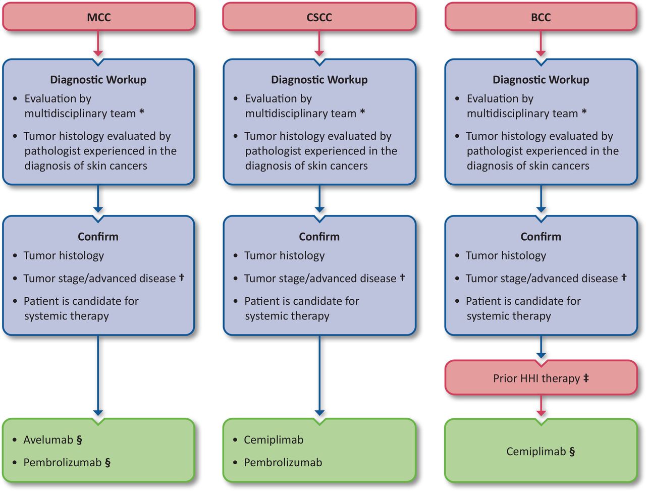
Compared with first-line chemotherapy, responses to ICIs have been found to be more durable and offer prolonged survival for a subset of patients with advanced MCC.52 Two ICIs have received FDA approval for the treatment of MCC at the time of guideline publication: avelumab and pembrolizumab.8 A summary of the FDA-approved ICIs (at the time of guideline publication) for MCC, CSCC, and BCC is depicted in figure 1.

FDA-approved ICI agents for NMSCs. Whenever possible, patients should be offered participation in clinical trials. Algorithm is intended to provide guidance and should not supplant sound clinical judgment—recommendations should be applied if feasible and as appropriate for individual patients. See product package inserts and the Approved anti-PD-(L)1 agents for MCC, Approved anti-PD-1 agents for CSCC, and Approved immunotherapy agents for BCC sections for more information on specific indications. *Some patients with advanced NMSC will be eligible for tissue-agnostic indications based on TMB and MSI/dMMR status. See the Tissue-agnostic indications for ICIs section for more information. †Advanced disease is defined in this guideline as tumors that are locally advanced, recurrent, and/or metastatic and not amenable to curative surgery or radiotherapy (box 1). ‡Or for whom an HHI is not appropriate. §Accelerated approvals contingent on confirmatory trials at the time of guideline publication. BCC, basal cell carcinoma; CSCC, cutaneous squamous cell carcinoma; dMMR, mismatch repair deficient; FDA, US Food and Drug Administration; HHI, hedgehog pathway inhibitor; ICI, immune checkpoint inhibitor; MCC, Merkel cell carcinoma; MSI, microsatellite instability; NMSC, nonmelanoma skin cancer; TMB, tumor mutational burden.
The landmark clinical trial outcomes leading to approvals for ICI indications for MCC are summarized in table 2. There is no evidence of clinical benefit thus far to favor one ICI over the other. Differences in reported response rates may be attributable to differing patient selection across trials.52 Additionally, as discussed in the Immune biomarkers for response to ICIs for MCC section, both the JAVELIN Merkel 200 and KEYNOTE-017 studies performed exploratory studies on various biomarkers including tumor virus status, PD-L1 expression, tumor-infiltrating lymphocyte (TIL) densities, and TMB. While several biomarkers have been associated with trends toward improved objective response rate (ORR), progression-free survival (PFS), and OS, no requirements for companion diagnostic testing were identified in the label indications due to low predictive power.
Landmark clinical trial data for FDA-approved immunotherapies for MCC
Approved anti-PD-(L)1 agents for MCC
Avelumab became FDA-approved in March 2017 for the treatment of metastatic MCC.6 The accelerated approval was based on the phase II, open-label, international study, JAVELIN Merkel 200 (NCT02155647), which enrolled a total of 204 patients into two arms: part A included patients with metastatic MCC whose disease had progressed after first-line chemotherapy, and part B included patients with metastatic or distally recurrent MCC who had not undergone prior systemic treatment (with the exception of adjuvant chemotherapy given with no clinically detectable or metastatic disease ≥6 months prior to study start). Patients in both arms received avelumab at 10 mg/kg intravenously (IV) every 2 weeks until disease progression or intolerable adverse events (AEs) occurred, with ORR being the primary endpoint measured by Response Evaluation Criteria in Solid Tumors v1.1 (RECIST v1.1). Approval was based on efficacy results from 88 patients in part A after the final patient enrolled had completed 12 months of follow-up (table 2). At longer follow-up (median 40.8 months; range 36.4─49.7), the ORR was maintained, with complete response (CR) achieved in 10 patients (11.4%) and partial response (PR) in 19 (21.6%). The median duration of response (DOR) was 40.5 months (95% CI 18 to not estimable [NE]), and at ≥44 months follow-up (as of May 2019) the OS was 12.6 months (95% CI 7.5 to 17.1).53 Responses were seen early, with 20 of 29 patients having confirmed responses by week 7, and 27 with a response by week 13.54 A rapid response was associated with improved survival. The 20 patients with an objective response at 7 weeks had a significantly improved 18-month survival probability of 90% (95% CI 65.6% to 97.4%), compared with 26.2% (95% CI 15.7% to 37.8%) for those without a rapid response. Significant differential survival probabilities were also seen in patients with objective responses at 13 weeks. PFS rates of the 88 patients in part A at 1 year, 2 years, and 3 years were 30% (95% CI 21% to 41%),55 26% (95% CI 17% to 36%), and 21% (95% CI 12% to 32%),53 respectively. In addition, patients treated with avelumab have reported experiencing improved QOL associated with less disease progression.56
In part B of the JAVELIN Merkel 200 study, the primary analyses in 116 total patients with a median follow-up of 21.2 months (range 14.9─36.6) reported an ORR of 39.7% (table 2) with 16.4% (n=19) experiencing a CR and 23.3% (n=27) a PR.57 The study also reported a durable response rate, defined as a CR or PR lasting ≥6 months, of 30.2% (95% CI 22.0% to 39.4%). Responses were observed by 3 months in a majority of patients, with a median time to response of 6.1 weeks (range 5–36). The median PFS was 4.1 months (95% CI 1.4 to 6.1). Median OS was 20.3 months (95% CI 12.4 to NE), and 6- and 12-month OS rates were 75% (95% CI 66% to 82%) and 60% (95% CI 50% to 68%), respectively.
Safety results from JAVELIN Merkel 200 demonstrate avelumab to be generally tolerable for patients with MCC. At follow-up analyses of ≥36 months, treatment-related adverse events (TRAEs) of any grade were reported in 68 of the patients in the part A arm (77.3%), with 10 patients (11.4%) experiencing a TRAE of grade ≥3. Immune-related adverse events (irAEs) were documented in 19 patients (21.6%), with 4 patients (4.5%) experiencing grade ≥3 irAEs. Overall, 8 patients (9.1%) had to cease treatment due to TRAEs, and no treatment-related deaths occurred.53 Similar safety profiles were seen in the part B arm, where 94 out of 116 total patients (81%) had any-grade TRAEs; 20 patients (17.2%) experienced a grade 3 TRAE, 1 patient experienced a grade 4 TRAE (dermatitis psoriasiform), and treatment was discontinued in 14 (12.1%) patients.57 Toxicities determined to be irAEs were seen in 35 (30.2%) patients, and 7 (6%) experienced a grade ≥3 irAE.
In December 2018, pembrolizumab was granted accelerated approval by the FDA for the treatment of adult and pediatric patients with recurrent, locally advanced, or metastatic MCC.8 Approval was based on tumor response rates and DORs in the KEYNOTE-017 trial (NCT02267603), which enrolled 50 subjects with locally advanced and metastatic MCC to receive 2 mg/kg IV pembrolizumab every 3 weeks as their first systemic treatment. All patients enrolled had unresectable stage IIIB (14%, n=7) or stage IV (86%, n=43) disease. Tumor responses were measured by RECIST v1.1 criteria. At a median follow-up of 14.9 months (range 0.4─36.4+), the ORR was 56% (95% CI 41.3% to 70.0%), with 12 CRs (24%; 95% CI 13.1% to 38.2%) and 16 PRs (32%; 95% CI 19.5% to 46.7%),58 and at a longer median follow-up of 31.8 months, the ORR was 58% (95% CI 43.2% to 71.8%).59 Responses were both rapid and durable; the median time to response was 2.8 months (range 1.5─9.7)58 and median DOR was not reached at 3 years (range 1.0+ to 51.8+ months).59 The 2- and 3- year OS rates were 68.7% and 59.4%, and PFS rates were 48.3% and 39.1%, respectively.58 59 Pembrolizumab was generally safe and well tolerated. TRAEs of any grade occurred in 96% of patients (n=48), with grade ≥ 3 experienced in 28% (n=14). Seven patients (14%) discontinued therapy due to TRAEs.58 A phase III confirmatory trial evaluating pembrolizumab for the treatment of patients with advanced MCC (KEYNOTE-913, NCT03783078) was ongoing at the time of guideline publication.
Locally advanced or metastatic MCC is often challenging to treat based on the anatomical location of disease, previous interventions, concomitant immunomodulation for autoimmune disease, a history of solid organ transplant, or the presence of hematological malignancies. Considerations for these challenging-to-treat populations are discussed further in the Patient selection for immunotherapy treatment of MCC section, and duration of treatment as well as surveillance are discussed in the Response monitoring and surveillance section. Generally, a multidisciplinary approach to care is optimal, with consideration of referral to an academic or high-volume center for best outcomes in particularly challenging cases. Enrollment in clinical trials for patients with contraindications to on-label indications for ICIs or with refractory/resistant disease is also important as trial settings may provide patients with access to potentially life-saving care that would be impossible to obtain in routine practice, in addition to continuing to make progress in identifying additional options in this heretofore largely untreatable disease60 61 (for more discussion of options for ICI-refractory or ICI-resistant disease, see the Anti-PD-(L)1 resistance section).
Immune biomarkers for response to ICIs for MCC
At the time of guideline publication, viral status does not have an established clinical utility for predicting benefit with ICIs. Neither KEYNOTE-017 nor JAVELIN Merkel 200 found a significant association between response rates to ICIs and MCPyV viral status. In KEYNOTE-017, the ORRs with pembrolizumab for patients with MCPyV-positive and MCPyV-negative tumors were 59% (19 of 32 patients) and 53% (9 of 17 patients), respectively (p=0.765).58 In cohort A (patients with metastatic MCC with disease progression after at least one prior line of chemotherapy) of JAVELIN Merkel 200, ORRs with avelumab in patients with MCPyV-positive tumors were 28.3% (95% CI 16.0% to 43.5%; n=46) and 35.5% (95% CI 19.2% to 54.6%; n=31) for patients with MCPyV-negative tumors.53 A similar trend was seen in cohort B (patients with metastatic MCC that was chemotherapy-naïve).57
Other immune-related biomarkers with well-characterized predictive power for benefit with checkpoint blockade in some solid tumor types include PD-L1 expression on tumor cells (TCs) and/or immune cells (ICs), high tumor mutational burden (TMB-H), high microsatellite instability (MSI-H), and IC infiltration. Although several of these immune biomarkers have been included in exploratory analyses, currently, no validated biomarkers exist to predict response to ICIs for patients with MCC. Efforts are ongoing, however, to develop reliable multifactor prediction tools.
PD-L1 expression in the tumor microenvironment has been correlated with increased response rates in multiple disease settings, and confirmed ‘positivity’ by an approved companion diagnostic is indicated for on-label use of ICIs in some cancers, such as breast cancer, lung cancer, urothelial cancer, and others.8 62 63 MCC is generally considered an immunologically inflamed tumor, with frequent PD-L1 expression and IC infiltration. One analysis reported PD-L1 positivity rates of 49% for TCs and 55% for ICs in MCC diagnostic pathology specimens across stages.17
In the registrational trials for ICIs in MCC, no association was seen between tumor PD-L1 expression and ORRs. In KEYNOTE-017, pretreatment PD-L1 expression was assayed by IHC using the anti-PD-L1 22C3 antibody, and ‘PD-L1-positive’ tumors were defined as tumors with positive PD-L1 staining on ≥1% of TCs or ICs. Trends were observed toward increased OS (p=0.057) and PFS (p=0.128) in patients with >1% PD-L1 TC expression; however, these analyses were exploratory in nature.58 The JAVELIN Merkel 200 trial also found that ORR was not significantly different between patients with PD-L1-positive (PD-L1 expression in ≥1% of TCs) and PD-L1-negative tumors, as scored by a proprietary PD-L1 IHC assay (using the 73-10 anti-PD-L1 antibody), although similar trends for better response and OS rates were seen in the small numbers of patients who had PD-L1-positive tumors. In cohort A of the study (patients with metastatic MCC with disease progression after at least one prior line of chemotherapy), of the 22 patients with long-term OS (defined as >36 months) after treatment, 81.8% had PD-L1-positive tumors, whereas only 18.2% had PD-L1-negative tumors.53 Although the study was not powered to compare PD-L1 expression subgroups, the data suggest that PD-L1 expression may be more prognostic than predictive in MCC. Further research is needed to identify and validate biomarkers for immunotherapy efficacy in MCC.
Several of the registrational trials for ICIs in MCC have included translational studies on tumor-infiltrating ICs,53 57 64–66 which generally portend a more favorable prognosis with higher levels of IC infiltration. However, these measures are not yet validated or readily available as predictive biomarkers for response.
Because the main etiological factors underlying tumorigenesis are either exposure to UV radiation or MCPyV infection, the mutational landscapes of MCC tumors, although heterogeneous, tend to cluster around two distinct signatures20 67: TMB-H MCC tumors (characteristic of UV exposure) tend not to harbor MCPyV genomes and vice versa. The mutation frequencies of MCPyV-positive tumors have been reported as 20- to 90-fold lower than for MCPyV-negative tumors.67 68 The mutational landscape of MCPyV-negative tumors shows strong UV signatures.20 68 69 TMB-H has also been correlated with improved response to immunotherapies in multiple types of solid tumors70 and is an approved tissue-agnostic eligibility criterion for treatment with pembrolizumab monotherapy8 based on improved ORR and DOR in patients with tumors harboring 10 or more mutations/megabase (Mb) in KEYNOTE-158 (for further discussion, see the Tissue-agnostic indications for ICIs section).71 The role of TMB as a predictor for efficacy is thought to be, in part, due to increased neoantigen presentation eliciting antitumor cytotoxic T cell responses.11 In MCC, however, data are still lacking on whether TMB-H predicts improved outcomes with ICI therapy, although trends for numerically higher responses have been reported.53 57 Despite the distinct genomic landscapes associated with individual etiologies, outcomes with ICIs have been similar across viral and non-viral MCCs in published trials.20 53 The published cohorts to date have been small, however, and further research is needed to understand determinants of response to ICIs in MCC.
Panel recommendations
For patients with metastatic, recurrent, or locally advanced MCC that is not amenable to curative surgery or radiation and for whom no contraindications to immunotherapy are present, first-line therapy with an approved anti-PD-(L)1 ICI is recommended (LE:2).
For patients with MCC who experience disease progression while on anti-PD-(L)1 immunotherapy, therapeutic options are limited and include clinical trials or chemotherapy. Switching treatments from one anti-PD-(L)1 antibody to another anti-PD-(L)1 antibody is unlikely to be beneficial.
While responses to PD-(L)1 blockade are frequent and generally durable, disease progression may occur in a subset of responders, and hence, continued surveillance for MCC progression is warranted.
For patients with MCC being treated with anti-PD-(L)1 ICIs, there are no validated biomarkers to predict benefit (including MCPyV viral status or PD-L1 expression).
Novel strategies and promising future directions for MCC
Several strategies are under investigation to expand the population of patients that benefit with immunotherapy, including neoadjuvant and/or adjuvant ICIs, combination approaches, novel ICIs, cytokine therapies, agonists of immune-related receptors, intratumoral therapies, and cellular therapies. Because MCC is a rare disease, efficacy for novel agents is often evaluated in basket trials that include patients with multiple tumor types, including other skin cancers or virus-associated solid malignancies.
Investigational immunotherapies for MCC in the neoadjuvant and adjuvant settings
The risk of recurrence in MCC is high, and thus, neoadjuvant and adjuvant therapies have been active areas of study, with adjuvant cytotoxic chemotherapy showing no discernable survival benefit at any stage of disease.72 73 The benefit of immunotherapy in earlier stages of MCC has not yet been established. Importantly, despite favorable reports of safety and efficacy, the potential for disease progression during neoadjuvant therapy, possibly to the point where the tumor becomes unresectable, is a major concern.
CheckMate 358 (NCT02488759), a multicohort phase I/II trial, demonstrated a pathologic complete response (pCR) rate of 47.2% (17 of 36) in patients with MCC who received 2 cycles of nivolumab followed by surgery.74 Median recurrence-free survival (RFS) and OS had not been reached at a median follow-up of 20.3 months. The safety profile was acceptable, with TRAEs occurring in 46.2% of patients and grade 3–4 TRAEs in 7.7% of patients. Three patients (7.7%) were unable to undergo surgery due to disease progression (n=1) or AEs (n=2). Further studies including comparator groups with adjuvant ICIs and resection alone are needed to understand if neoadjuvant therapy improves outcomes for patients with resectable MCC. Clinical trials are the most appropriate context for these approaches at this time.
Several trials are ongoing to investigate whether ICIs used in the adjuvant setting can reduce the risk of recurrence. No improvement in disease-free survival (DFS) (HR 1.8; 95% CI 0.3 to 10; p=0.48) and significantly increased incidence in AEs was seen in 40 patients with completely resected MCC who were treated with adjuvant anti-CTLA-4 ipilimumab in the ADMEC-O study (NCT02196961).75 An additional arm of ADMEC-O investigating adjuvant nivolumab for completely resected MCC tumors is ongoing (NCT02196961). Additional trials are comparing avelumab as an adjuvant therapy against placebo, such as the phase III ADAM trial (NCT03271372) and the phase II I-MAT trial (NCT04291885). Finally, several studies are combining adjuvant radiation therapy with ICIs, including the phase III STAMP study (NCT03712605), which is evaluating pembrolizumab plus optional radiation therapy versus observation plus optional radiation therapy after complete resection in patients with stage I–III MCC. Importantly, however, the risks of adjuvant therapy include increased AEs and treatment-related mortality, and there is no direct evidence to support the routine use of radiation with ICIs outside of a clinical trial setting at this time.
Investigational immunotherapies for MCC in the advanced/metastatic setting
The incorporation of the approved anti-PD-(L)1 ICIs into the standard of care has improved outcomes for patients with advanced unresectable MCC. Building on these early successes, studies are under way to investigate additional immunotherapies for advanced unresectable MCC.
While avelumab and pembrolizumab are the only FDA-approved ICIs for the treatment of MCC at the time of guideline development, ongoing studies are evaluating other anti-PD-(L)1 agents. For example, early results (median follow-up of 26 weeks [range 5─35]) from the phase I/II CheckMate 358 trial evaluating systemic nivolumab for the treatment of advanced virus-associated tumors after two or fewer prior therapies, found an ORR of 68% (95% CI 48% to 86%; n=15) among the 22 patients with MCC, with ongoing responses in 13 patients.76 Although some guidelines recommend nivolumab or other anti-PD-(L)1 ICIs for advanced MCC,27 their use is off-label at the time of guideline preparation. Based on the known mechanisms of anti-PD-(L)1 therapies and response rates in published trials, however, there is no indication that nivolumab or other off-label anti-PD-(L)1 agents differs substantially from approved agents in efficacy and tolerability.
Several studies are investigating immune-modulatory agents targeting the innate and adaptive immune systems, with and without ICIs as a combination regimen, to enhance antitumor responses. Some of these strategies include local interleukin (IL)-12, IL-15 superagonists, IL-17, oncolytic viruses, radiation therapy, and various toll-like receptor (TLR) agonists.
Immune evasion mechanisms have been demonstrated to be active in patients with MCPyV-positive MCC tumors, including MCPyV-specific CD8+ T cell dysfunction due to high expression of the inhibitory receptors PD-1 and T cell immunoglobulin and mucin domain-containing protein 3 (TIM-3),77 and downregulation of class I human leukocyte antigen (HLA-I).78 To overcome these endogenous mechanisms, several clinical trials are using adoptive cell therapies to expand autologous MCPyV-specific cytotoxic T cells to treat patients in combination with recombinant interferon-β (IFN-β) and radiation therapy,79 80 both shown to upregulate HLA-I expression.78 81 Several allogenic cell therapies are also being studied using both virus-specific and other IC transfers.
Treatment options for patients with MCC whose disease has progressed on prior anti-PD-(L)1 therapies (discussed further in the Anti-PD-(L)1 resistance section) are also being studied. Of note, trials under way include the CARTA study (NCT04792073) assessing comprehensive ablative radiation therapy with or without avelumab, and the DUET-1; 02 study (NCT04590781) using a bispecific somatostatin receptor 2 (SSTR2) and CD3 binding antibody, tidutamab. In addition, two retrospective studies that treated patients’ MCC with salvage anti-PD-1 nivolumab in combination with anti-CTLA-4 ipilimumab in patients who have progressed on prior anti-PD-(L)1 therapy have reported responses in 3 of 582 and 4 of 1383 patients. Additionally, clinical trials assessing cell therapy combinations for patients with anti-PD-(L)1 refractory disease are also under way, though most are in early phases (NCT03747484).
Panel recommendations
For patients with resectable MCC at high risk of recurrence, enrollment in clinical trials of neoadjuvant or adjuvant therapy should be offered where available.
For patients with unresectable, locally advanced, metastatic, or recurrent MCC, enrollment in clinical trials of novel agents and novel combinations should be offered where available.
Patient selection for immunotherapy treatment of MCC
Immunotherapy should be considered within the broader context of available therapies for MCC. Both of the currently FDA-approved immunotherapies for MCC, avelumab and pembrolizumab, are indicated specifically for patients with advanced forms of the disease.6 8
For those patients with unresectable tumors or with contraindications to surgery or radiation, systemic therapy should be considered. Reasons to avoid surgery and/or radiation include inability to achieve resection with microscopically negative margins, medically unfit patients for surgical or radiation intervention, or treatment-related morbidity that would significantly impact the patient’s QOL. Of the systemic therapy options, chemotherapy (as discussed in the Recommended immunotherapies for MCC section) has not demonstrated durable benefit as an initial treatment for advanced MCC, with high initial response rates but poor PFS,84 nor as an adjuvant therapy at any disease stage of MCC.73 ICIs have become the new standard for patients with advanced MCC, often with long-lasting responses.
Special patient populations for MCC
In addition to the general label indications to determine which patients with MCC are candidates for the approved ICIs, special considerations need to be taken for certain patient populations, especially those with altered or suppressed immune systems at baseline. Patients with HIV have typically been excluded from clinical trials of ICIs due to a lack of data on how ICIs would interact with altered immune systems. This is an important population to consider, however, as HIV-positive individuals are at elevated risk of developing MCC, presumably due to their immunocompromised state.85 Although data on the safety of ICIs specifically in HIV-positive patients with MCC are lacking, a systematic review of 73 patients with HIV and a variety of solid tumors demonstrated that ICIs were well tolerated and efficacious, with ORRs and frequencies of grade 3–4 irAEs similar to those found in patients without HIV. Furthermore, of 28 patients with no detectable HIV load prior to ICI therapy, HIV remained controlled in 26 (93%) after treatment with checkpoint inhibition.86 Overall, patients with HIV can be treated with ICIs, ideally with the viral status well controlled (ie, CD4 count ≥100 cells/µL, HIV viral load <200 copies/mL, and on antiretroviral therapy), however, treatment for patients with viral status that does not meet these parameters can still be considered.
Other patient populations that have frequently been excluded from clinical trials of ICIs include patients with pre-existing autoimmune disease and solid organ transplant recipients, due to the possibility that ICI-mediated immune activation could lead to flares of autoimmunity or graft rejection, respectively. A retrospective study of 30 patients with melanoma and an existing autoimmune disease treated with ipilimumab found an ORR of 20%, with grade 3–5 irAEs in 33% of patients. Although 27% of patients experienced a flare of their autoimmune disease, all of the flares were manageable with corticosteroids.87 A subsequent systematic review that included 123 patients with pre-existing autoimmune disease and a variety of tumor types treated with anti-PD-(L)1 and/or anti-CTLA-4 ICIs found that 41% of patients had an exacerbation of their autoimmune disease, 25% developed de novo irAEs, and 9% experienced both.88
ICI therapy administered to solid organ transplant recipients may be efficacious but carries a high risk of allograft rejection, as demonstrated in retrospective analyses. One report describing 39 solid organ transplant recipients with various solid tumors who received ICIs found that 41% (n=16) experienced allograft rejection, leading to graft loss in 81% (n=13). Among the 22 patients with melanoma and 5 with CSCC in the study, allograft rejection occurred in 8 patients with melanoma and 2 patients with CSCC. Favorable antitumor responses were seen in 36% of patients with melanoma, and all patients with CSCC achieved a CR or a PR.89 A more recent pooled analysis described similar results, with a rate of allograft rejection at 41% in a total of 64 patients.90 Currently, NMSCs such as MCC lack tumor-specific data on the risk of graft rejection and successful management strategies. Therefore, ongoing and future clinical trials, such as NCT03816332, which is investigating the use of ICIs in combination with standardized transplant immunosuppression in kidney transplant recipients diagnosed with unresectable or metastatic cutaneous malignancies, will be critical in better understanding how to optimize the safety and efficacy of immunotherapy in these patient populations.
Patients with chronic lymphocytic leukemia (CLL) are immunocompromised in addition to being at increased risk of developing MCC.91 Patients with some NMSC and concurrent hematological malignancies may have worse outcomes with ICI treatment;92 however, prospective data are lacking at this time and trials are ongoing. A small number of reports have demonstrated general safety using ICIs alone or in combination with ibrutinib.93 94
Some patients who are being considered for immunotherapy are on immunosuppressive drugs, such as corticosteroids, to treat pre-existing autoimmune conditions. Corticosteroids suppress the immune system, and steroid exposure is independently associated with worse survival outcomes in advanced solid tumors.95 However, corticosteroids are also a cornerstone of irAE management, and studies to date have not conclusively demonstrated that short-term (ie, limited duration for the purposes of toxicity management and promptly weaned when AEs resolve) immunosuppression affects clinical benefit with ICI therapy. A systematic review of 4,045 patients with a variety of tumor types found that patients taking high-dose steroids (>10 mg prednisone or equivalent) were at increased risk of death or disease progression compared with patients who were not. This increased risk was greatest in subgroups of patients who were taking steroids for supportive care or for brain metastases. Notably, patients who received steroids to treat irAEs stemming from ICI therapy did not exhibit reduced OS.96 Similar results were found in a study of patients with non-small-cell lung cancer, where patients who received ≥10 mg prednisone at the time of beginning immunotherapy had worse median OS and PFS if receiving the prednisone for palliative care, but those receiving ≥10 mg prednisone for cancer-unrelated reasons (such as prior autoimmune conditions) experienced OS and PFS similar to those who received <10 mg prednisone.97 While these studies demonstrate that the context of steroid use is important, in general, for patients with immunological comorbidities (other than solid organ transplant preservation) who are taking high-dose corticosteroids and/or other immunosuppresives, a reduction to the minimum necessary dose is best practice prior to treatment with ICIs, and enrollment in clinical trials is encouraged if available. A skin cancer multidisciplinary tumor board or referral to a tertiary care center with a tumor board for evaluation of clinical trial eligibility may be beneficial for these difficult-to-treat patient populations.
Currently, there are limited data on the safety of pregnancy and ICI therapies (currently considered category D by FDA for pregnancy). In a review of available case studies (n=7) on the safety of ICI use in pregnant patients with melanoma, complications were observed in 71.4% of patients, and the average gestational age of delivery (n=9) was 30.4 weeks (range 24–38).98 In addition to considering the effects of ICIs on pregnancy, it is also important to consider the potential need for immunosuppressants for the treatment of irAEs, some of which, such as mycophenolate mofetil, have been shown to cause fetal malformation in the context of solid organ transplant.99 100 In addition, data are lacking on the effects of ICIs on fertility in women of childbearing age. Because endocrine irAEs may arise and persist long term after cessation of therapy, a referral to an onco-fertility specialist may be considered.
Panel recommendations
For solid organ transplant recipients with MCC who are potential candidates for ICI therapy, healthcare professionals should have a careful risk–benefit discussion with the patient that includes the potential risk of allograft loss, which may result in the need for dialysis (ie, for kidney transplant recipients) or death (eg, recipients of heart, lung, or liver transplants) (LE:1). Chemotherapy is an acceptable alternative first-line therapy in this population (LE:3). Enrollment in clinical trials should be encouraged.
For patients with MCC on therapeutic immune suppression, a discussion should be initiated on reducing or modifying immune suppression, if appropriate, before proceeding with ICI therapy.
For patients with MCC who are receiving high-dose corticosteroids for reasons other than solid organ allograft preservation, the dose of corticosteroids should be reduced to ≤10 mg prednisone (or equivalent) per day, if possible, prior to initiation of ICI therapy.
Caution is advised when treating with ICIs in patients with MCC who have a history of autoimmune disease, as they may experience a flare of their autoimmune disease and/or a distinct irAE. Healthcare professionals should have a careful risk–benefit discussion with the patient, which includes the potential risks associated with an autoimmune flare (LE:1).
Cutaneous squamous cell carcinoma
CSCC is an extremely common form of skin cancer.4 The vast majority of CSCCs are small, local tumors with an excellent prognosis and are treated with surgical excision alone. Few options exist, however, for the small subset of patients with locally advanced or metastatic disease. For tumors that are not amenable for surgery or radiation, two ICIs, cemiplimab and pembrolizumab, have been approved by the FDA for the treatment of CSCC (discussed in the Recommended immunotherapies for CSCC section).
For the purpose of this article and the expert panel recommendations, ‘advanced CSCC’ is defined as tumors that are not eligible for curative surgery or radiation, recurrent, and/or metastatic (box 1). In the case of advanced CSCC, multidisciplinary care team management is optimal and may include surgical oncology, radiation oncology, medical oncology, dermatology, and any other pertinent specialties, as appropriate.
Diagnosis, workup, and initial staging for CSCC
As with any cancer, accurately differentiating CSCC from other types of skin cancer is crucial for appropriate clinical care. The gold standard diagnostic test is H&E analysis of a tumor biopsy specimen, which can distinguish in situ CSCC, invasive CSCC, and histological variants.101 When CSCC tumors are poorly differentiated and/or when the origin of the tumor is in question, IHC is currently the most useful technique to confirm the diagnosis. Positive IHC staining for p63 or its isoforms lacking a transactivation domain (stained with anti-p63 and anti-p40 antibodies, respectively) can aid in confirming CSCC.102 103 Staining for cytokeratins can also be used for confirming a CSCC diagnosis, in addition to distinguishing histological variants of CSCC.104
In addition to IHC, tumor genetic profiling by NGS in select cases can support the diagnosis or management decisions for CSCC, which, like virus-negative MCC, is highly mutated compared with other solid tumors. A common risk factor for CSCC is exposure to UV radiation,105 and the mutational landscape of CSCC tumors commonly shows a high mutational burden with a UV signature,106 which can support the diagnosis of CSCC. A recent study found the median somatic mutation rate of CSCC to be 61.2 mutations/Mb (n=39), whereas melanoma, head and neck squamous cell carcinoma, and lung squamous cell carcinoma have reported median mutation rates of 13.2, 3.2, and 8.2 mutations/Mb, respectively.106
Two systems are primarily used for staging of primary CSCCs: the 8th edition of AJCC TNM staging system (which applies to the head and neck region only)39 and the Brigham and Women’s Hospital (BWH) T staging system, which assesses primary tumors but does not address lymph nodes.107 BWH T staging has been demonstrated to have higher specificity and better positive predictive value in identifying the likelihood of metastasis or death compared with the AJCC 8th Edition staging, but the two systems do not differ in their ability to predict OS or local recurrence.108 Summaries of the AJCC 8th Edition TNM and BWH T staging systems are provided in table 3 and table 4, respectively.
Summary of AJCC (8th Edition) TNM staging for CSCC* (adapted from Cañueto and Román-Curto176)
BWH T staging system for CSCC (adapted from Ruiz et al108)
The high-risk features that are included in the various staging systems for CSCC include tumor size, depth of invasion, histological differentiation, and perineural invasion of large caliber nerves. Histological LVI and immune-compromised status are considered high-risk characteristics but are not included in the staging systems. The number of these features that are positive will inform the stage of disease and could inform treatment decisions.101 109 For patients with advanced CSCC being considered for systemic immunotherapy, appropriate radiological imaging should also be performed to assess for metastatic disease.
Panel recommendations
Histopathological diagnosis of advanced CSCC should include H&E evaluation by a pathologist with experience in the diagnosis of skin cancers, when feasible.
For patients with advanced CSCC, multidisciplinary care team management is optimal.
Patients with CSCC who are being considered for systemic therapy should be evaluated by appropriate radiological imaging.
Recommended immunotherapies for CSCC
Prior to the approval of ICIs, there was no consensus on standard of care systemic therapies for metastatic and locally advanced CSCC not amenable to surgery or radiation. The most commonly used systemic treatments were targeted epidermal growth factor receptor (EGFR) inhibitors, cytotoxic platinum-based chemotherapies, and the antimetabolite 5-fluorouracil. The rationale for the use of these agents in CSCC was largely extrapolated from the treatment of mucosal head and neck squamous cell cancers. The advent of immunotherapy and subsequent rapid widespread adoption of ICIs, coupled with the recognition that tumors with very high TMB tend to be responsive to checkpoint blockade, motivated the evaluation and subsequent approval of two anti-PD-1 therapies in CSCC.
Approved anti-PD-1 agents for CSCC
Two anti-PD-1 ICIs have been approved by the FDA for the treatment of CSCC: pembrolizumab and cemiplimab (figure 1). Key outcomes from the landmark trials leading to the FDA-approved ICI indications for CSCC are summarized in table 5. Importantly, there is no evidence of clinical benefit thus far to favor one ICI over the other.
Landmark clinical trial data for FDA-approved immunotherapies for CSCC
In September 2018, the FDA approved cemiplimab for the treatment of patients with metastatic or locally advanced CSCC who are not candidates for curative surgery or radiation.7 Cemiplimab was the first approved ICI for CSCC, with a regulatory decision based on data from the phase I dose-escalation Study 1423 (NCT02383212) and the ongoing phase II open-label EMPOWER CSCC 1/Study 1540 (NCT02760498). The trials initially enrolled a total of 108 patients with metastatic, recurrent, or locally advanced CSCC who were not candidates for curative surgery or radiation for cemiplimab treatment at 3 mg/kg IV every 2 weeks (an additional 56-patient cohort received cemiplimab at 350 mg IV every 3 weeks [the approved dosage]). Patients in both trials had commonly received prior surgery (96% of patients) or radiotherapy (79% of patients), and 50% of patients had received prior systemic therapy. The approval was based on ORR and DOR data at a median follow-up of 8.9 months in the combined metastatic and locally advanced cohorts.
Follow-up analyses of EMPOWER CSCC 1/Study 1540 found an ORR of 50.8% (95% CI 37.5% to 64.1%) with a median duration of follow-up of 18.5 months in the metastatic cohort, an ORR of 44.9% (95% CI 33.6% to 56.6%) with a median duration of follow-up of 15.5 months in the locally advanced cohort, and an ORR of 42.9% (95% CI 29.7% to 56.8%) with a median follow-up of 17.3 months in the fixed dosage cohort, for a combined ORR from all three groups (n=193) of 46.1% (95% CI 38.9% to 53.4%, median duration of follow-up 15.7 months).110 CRs were achieved in 16.1% of patients, and PRs in 30.1% of patients. The median observed time to response across cohorts was 2.1 months (IQR 1.9–3.7). Although median DORs were not reached at the time of reporting, the estimated rates of patients with observed DORs of ≥12 months and ≥24 months were 89.5% and 68.8% in the metastatic cohort and 83.2% and 62.5% in the locally advanced cohort, respectively.
The most recent safety data available at the time of guideline publication reported that grade ≥3 TRAEs occurred in 17.1% of patients.110 Sponsor identified irAEs were reported in 29.5% of patients and at least one grade ≥3 irAE in 9.3%.
Pembrolizumab was approved by the FDA in June 2020 for the treatment of patients with recurrent or metastatic CSCC not curable by surgery or radiation.8 Approval was based on ORR and DOR in the phase II, open-label KEYNOTE-629 trial (NCT03284424). Of the 105 patients enrolled and treated with pembrolizumab at 200 mg IV every 3 weeks for up to a maximum of 24 months, 74.3% had prior radiation therapy, 86.7% had previously received at least one systemic therapy, and only 13.3% were receiving pembrolizumab as their first line of treatment.111 With an encouraging ORR reported at the time of approval (median follow-up of 9.5 months, table 5), the median DOR was not reached and 69% of patients had a median DOR of ≥6 months. In July 2021, the approval for pembrolizumab was expanded to also include patients with locally advanced CSCC not curable by surgery or radiation, based on an interim analysis of this cohort of patients in KEYNOTE-629.8
In an interim analysis of KEYNOTE-629 with longer follow-up (14.9 months [IQR 12.6–17.2] for the locally advanced cohort and 27.2 months [IQR 25.6─29.2] for the recurrent/metastatic cohort),112 the ORR for patients with locally advanced CSCC was 50.0% (95% CI 36.1% to 63.9%) with a CR rate of 16.7% and a PR rate of 33.3%. Median time to response was 2.6 months (IQR 1.4–3.6), with responses reported in 14 of the 27 responders by week 6. Median PFS and median DOR were not reached with an estimated 88.1% and 84.1% of responders having responses that lasted at least 6 and 12 months, respectively. Of the patients with baseline and postbaseline imaging available (n=48), 83.3% had a reduction in target lesion size, including 66.7% with at least a 30% reduction. Median OS was not reached, and the estimated OS rate at 18 months was 73.6% (95% CI 59.5% to 83.4%).
In the recurrent/metastatic cohort of KEYNOTE-629, the ORR was 35.2% (95% CI 26.2% to 45.2%), with a CR rate of 10.5% and a PR rate of 24.8%. The ORR was higher in patients who received pembrolizumab as first-line therapy (n=14) compared with patients who received pembrolizumab as second-line or later therapy (n=91; 50.0% vs 33.0%, respectively). The overall median time to response was 1.6 months (IQR 1.4–13.1), with responses reported in 20 of the 37 responders by week 6. Median PFS was 5.7 months (95% CI 3.1 to 8.5) and median DOR was not reached (95% CI 22.4 months to not reached) with estimates of 80.7% of responses lasting at least 6 months and 77.8% lasting at least 12 months. Among the 95 patients who had baseline and postbaseline evaluable images, 77.9% demonstrated a reduction in target lesion size, and 58.9% had at least a 30% reduction in target lesion size. Median OS was 23.8 months (95% CI 13.4 to 29.8), and the OS rates at 12 and 24 months were 61.0% (95% CI 50.9% to 69.5%) and 48.4% (95% CI 38.5% to 57.6%), respectively. Biomarker data from this study reported response to pembrolizumab occurred independent of PD-L1 status of tumors (discussed in the Immune biomarkers for response to ICIs for CSCC section). Additionally, the phase II, open-label CARSKIN trial (NCT02883556) is examining the efficacy and safety of pembrolizumab as a first-line therapy for unresectable locally or regionally advanced or metastatic CSCC. Results from 39 patients found a median PFS of 6.7 months and median OS was 25.3 months (95% CI 14.2 months to NE) at a median follow-up of 22.4 months. At 15 weeks, the response rate was 41% (95% CI 26% to 58%), with 3 CRs and 13 PRs. TRAEs occurred in 71% of patients, and 7% of patients experienced serious TRAEs.113
Pembrolizumab was found to be generally tolerable in KEYNOTE-629. TRAEs were observed in 69.2% of patients, with 11.9% experiencing grade ≥3 and 8.8% requiring a discontinuation of treatment. Infusion reactions or irAEs occurred in 22.6% of patients and were mostly grade ≤2, but 8.2% of patients experienced a grade ≥3 irAE or infusion reaction.112
While there are differences in the patient populations evaluated in the registrational studies (described in table 5), either cemiplimab or pembrolizumab is appropriate first-line therapy for patients with metastatic or locally advanced CSCC who are not candidates for curative surgery or radiation. In the published trials, tumor regression typically occurred within the first 2 months, but in some cases, delayed responses can occur and it may be appropriate to continue treatment if the patient is clinically stable and experiencing clinical benefit. Duration of therapy in both studies was not longer than 2 years, by design, and optimal duration of treatment is not known.
Similar to MCC, patients with locally advanced or metastatic CSCC may present with comorbidities that complicate an immunotherapy treatment plan. Considerations for challenging populations are discussed in the Patient selection for immunotherapy treatment of CSCC section, and duration of treatment and surveillance are discussed in the Response monitoring and surveillance section. Generally, a multidisciplinary approach to care is essential, with consideration for referral to an academic or high-volume center for best outcomes in particularly challenging cases. Enrollment in clinical trials for patients with contraindications or with refractory/resistant disease to ICI treatment is also important (see the Anti-PD-(L)1 resistance section)—not only because a trial setting may provide patients with access to potentially life-saving care that would be impossible to obtain in routine practice, but also to continue to make progress in identifying additional options in this heretofore largely untreatable disease.60 61
Immune biomarkers for response to ICIs for CSCC
Currently, there are no validated biomarkers to predict immunotherapy-related treatment response or inform clinical decisions for patients with CSCC, including PD-L1 expression or TMB. Tumor PD-L1 expression did not significantly correlate with response to PD-1 blockade with pembrolizumab in KEYNOTE-629, although there was a trend towards better ORR in tumors with a PD-L1 combined positive score (CPS, the number of PD-L1 staining cells [TCs, lymphocytes, and macrophages] divided by the total number of viable TCs, multiplied by 100) ≥1 in the combined analysis of patients with metastatic and locally advanced CSCC.112 Patients with tumors with PD-L1 CPS ≥1 (n=115) had an ORR of 42.6% (95% CI 33.4% to 52.2%), whereas for those with PD-L1 CPS <1 tumors (n=15), the ORR was 20% (95% CI 4.3% to 48.1%). In a separate analysis of response rates stratified by tumor proportion score (TPS, the number of positive TCs divided by the total number of viable TCs, multiplied by 100), similar ORRs in the PD-L1-positive group were seen when the threshold for positivity was 50%. In addition, PD-L1 expression in CSCC has been shown to correlate with risk for metastasis.15 114 115 Overall, the evidence at this time indicates that individuals with PD-L1-negative tumors may still derive benefit from the approved immunotherapies, and therefore all patients eligible for systemic immunotherapy should receive an ICI regardless of PD-L1 expression.
The value of TMB as a predictive or prognostic biomarker for patients with CSCC is an ongoing area of study. CSCC tumors have among the highest levels of TMB in solid malignancies, with a damage pattern characterized by UV signature mutations. One study including patients with a variety of squamous cell carcinomas from several tissue types found that both cutaneous origin and increasing TMB were associated with better outcomes with anti-PD-(L)1 blockade (monotherapy or in combination regimens). The link between TMB and clinical benefit with immunotherapy for CSCC specifically was not statistically significant in multivariate analysis; however, these data are difficult to interpret due to the small sample size.116
Owing to the high TMB of CSCC, identifying individual driver genes for prognostic use has been a challenge, and single-gene tests have had little utility thus far. However, the use of gene expression panels to profile transcriptional activity in tumors is an ongoing area of research to attempt to identify predictive or prognostic signatures. A commercially available gene expression panel (DecisionDx-SCC) uses a panel of 40 genes (including six control loci) to stratify primary tumors into low-risk, high-risk, and highest-risk categories for prediction of metastasis. The assay has a reported positive predictive value of 60% for patients with class IIB (highest risk) disease117; however, there are no prospective studies of its clinical utility, and thus, its use is not recommended at this time.
Panel recommendations
For patients with metastatic, recurrent, or locally advanced CSCC that is not amenable to curative surgery or radiation and for whom no contraindications to immunotherapy are present, first-line therapy with an approved anti-PD-1 ICI is recommended (LE:2).
While responses to PD-1 blockade are frequent and generally durable, disease progression may eventually occur in a subset of responders, and hence continued surveillance for CSCC progression is warranted.
For patients with CSCC being treated with anti-PD-(L)1 ICIs, there are no validated biomarkers that predict benefit (including PD-L1 expression).
Novel strategies and promising future directions for CSCC
A number of immunotherapies are in development for the treatment of CSCC in the adjuvant/neoadjuvant setting as well as for advanced metastatic disease, including new indications or combinations of ICIs, oncolytic viruses, and integration of ICIs at earlier stages of disease through adjuvant or neoadjuvant strategies.
Investigational immunotherapies for CSCC in the neoadjuvant and adjuvant settings
For patients with resectable CSCC tumors, there are currently no approved immunotherapy agents for neoadjuvant or adjuvant treatment. At the time of guideline preparation, several trials are investigating neoadjuvant ICIs, both as monotherapy and in combination with other ICIs, for the treatment of CSCC. The considerations and risk–benefit analyses for neoadjuvant use of ICIs in CSCC include the possibility for undue delay in surgery to the point of becoming unresectable and increased toxicity.
Although not current practice, one strategy that has reported encouraging data from non-randomized studies is a short course of neoadjuvant PD-(L)1 blockade followed by curative surgery. In a pilot study (NCT03565783) including 20 patients with CSCC of the head and neck, two preoperative doses of cemiplimab led to 11 pCRs and 3 major pathological responses in the resection specimens. Inflamed tumor microenvironments and enrichment for CD8+ tumor-specific T cells were observed in samples from tumors with pCR. DSS, DFS, and OS rates at 12 months were 95% (95% CI 85.9% to 100%), 89.5% (95% CI 76.7% to 100%), and 95% (95% CI 85.9% to 100%), respectively.118 A larger clinical trial (NCT04154943) for patients with CSCC is in progress to validate these findings. Both cemiplimab and pembrolizumab are being investigated in phase III studies (NCT03969004 and NCT03833167, respectively) for efficacy as adjuvant treatments in patients with high-risk or locally advanced CSCC tumors that have undergone complete/gross macroscopic resection and postoperative radiation therapy. However, as results for NMSCs, including CSCC, have yet to be reported, administration of adjuvant immunotherapies should only be done within a clinical trial.
Investigational immunotherapies for CSCC in the advanced/metastatic setting
Additional anti-PD-1 monotherapy studies are also ongoing. Two phase II trials are also currently investigating nivolumab for patients with advanced CSCC, NCT04204837 and NCT03834233. In the latter study, CA209-9JC, a phase II, open-label trial evaluating nivolumab for the first-line treatment of advanced or metastatic CSCC, the best ORR was 54.5% (n=22), and the median DOR, PFS, and OS had not been reached at the time of follow-up. Grade ≥3 TRAEs were reported in 21% of patients, with one discontinuation due to toxicity.119
ICI combination strategies for the treatment of advanced CSCC that have reached early-phase clinical trials include ICIs combined with radiation therapy, IL-17, targeted therapies (such as EGFR and MEK inhibition), and oncolytic viruses. Early data have been reported on the intratumoral use of the oncolytic herpes simplex type-1 virus RP1, which is engineered to increase immunogenic responses via delivery of granulocyte-macrophage colony-stimulating factor (GM-CSF) to the tumor microenvironment as well as via a fusogenic GALV-GP R-protein that causes TCs to form large multinucleated syncytia inducing cytotoxicity and promoting antigen presentation by dendritic cells. The agent has advanced through phase II trials for solid tumors, including CSCC. In the phase II IGNYTE trial (NCT03767348) of RP1 in combination with nivolumab that enrolled patients with either melanoma or NMSCs, interim results reported that 5 of 6 patients with CSCC responded within 1─7 months of treatment, with 3 achieving CR.120 RP1 is also being studied in combination with cemiplimab for treatment of advanced CSCC in the CERPASS study (NCT04050436).
Panel recommendations
For patients with resectable CSCC at high-risk of recurrence, enrollment in clinical trials of neoadjuvant or adjuvant therapy should be offered where available.
For patients with unresectable locally advanced or metastatic CSCC, enrollment in clinical trials of novel agents and novel combinations should be offered where available.
Patient selection for immunotherapy treatment of CSCC
For patients with CSCC who have contraindications to surgery and/or radiation, including inability to achieve resection with microscopically negative margins, being medically unfit for surgery or radiation, or having a treatment-related morbidity that would significantly impact the patient’s QOL, systemic therapies can be used. Although ICIs are generally well tolerated in most patients, the mechanisms behind the therapeutic benefit for ICIs also predicate a need for special considerations with some patient populations prior to use.
Special patient populations for CSCC
Similar to MCC, individuals who are immunosuppressed are more likely to develop CSCC,121 122 including those who have received solid organ transplants121 123 (who are more likely to develop aggressive CSCC),124 are HIV-positive,121 125 or have been diagnosed with CLL.126 These and other factors require special considerations for patients with CSCC when administering ICIs, including advanced patient age, high-dose corticosteroid use, and pre-existing autoimmune disease, which generally overlap with and are discussed in detail in the Special patient populations for MCC section. One phase II single arm study for immune-compromised patients—defined as having a history of HIV or a history of treated or active hematologic malignancies—with locally recurrent and/or metastatic CSCC (NCT04242173) is ongoing, evaluating cemiplimab. This study, and others, highlight the importance of collecting data on ICI safety and efficacy in patients considered ‘not typical’ candidates for immunotherapy, as well as providing access to care for these populations. Clinical trial enrollment is encouraged, if available, and remains a high priority for special patient populations.
Panel recommendations
For solid organ transplant recipients with CSCC who are potential candidates for ICI therapy, healthcare professionals should have a careful risk–benefit discussion with the patient that includes the potential risk of allograft loss, which may result in the need for dialysis (ie, for kidney transplant recipients) or death (eg, recipients of heart, lung, or liver transplants) (LE:1). Chemotherapy (LE:1) and/or EGFR inhibitors (LE:3) are acceptable alternative first-line therapies in this population. Enrollment in clinical trials should be encouraged.
For patients with CSCC who are receiving high-dose corticosteroids for reasons other than solid organ allograft preservation, the dose of corticosteroids should be reduced to ≤10 mg prednisone (or equivalent) per day, if possible, prior to initiation of ICI therapy.
Caution is advised when treating with ICIs in patients with CSCC who have a history of autoimmune disease, as they may experience a flare of their autoimmune disease. Healthcare professionals should have a careful risk–benefit discussion with the patient, which includes the potential risks associated with an autoimmune flare (LE:1).
For patients with CSCC on therapeutic immune suppression, a discussion should be initiated on reducing or modifying immune suppression, if appropriate, before proceeding with ICI therapy.
Immunotherapy for the treatment of other types of NMSCS
Other types of NMSCs besides MCC and CSCC include BCC, which is the most common type of NMSC, and cutaneous forms of angiosarcoma, Kaposi sarcoma, and adnexal tumors, among other rare skin neoplasms. Cemiplimab is currently approved for BCC, as is the topical immune modulator imiquimod, both of which are discussed in the Basal cell carcinoma section. Numerous studies are investigating immunotherapy for other skin malignancies, as discussed in the Immunotherapies in development for other NMSCs section. In addition, tissue-agnostic indications for ICIs may allow patients with rarer NMSCs to be treated on-label based on tumor features such as TMB-H or MSI-H, discussed in the Tissue-agnostic indications for ICIs section. Finally, as immunotherapies continue to be investigated for rarer NMSCs, off-label use of approved agents may be considered where appropriate. Caution should be taken for patients with Kaposi sarcoma and HIV, however, due to the potential risk of Kaposi sarcoma-associated herpesvirus-associated B-cell lymphoproliferation with ICI treatment.127 Clinical trial enrollment is always encouraged to provide access to care for patients and also move the field of immunotherapy forward, which is especially important in understudied rare cancers.
Basal cell carcinoma
BCC is the most common skin malignancy, with incidence rates on the rise.128 129 Most cases of BCC are localized and can be cured with surgical excision, with a very low rate of metastasis or recurrence; the overall incidence of metastasis ranges from 0.0028% to 0.55%.130 Similar to CSCC, the strongest environmental risk factors for BCC include UV radiation exposure and immunosuppression.131
Histological evaluation of localized tumors may identify variants at higher risk for recurrence.131 In addition to histological variants, large tumor diameter is also a predicting factor for BCC recurrence. One study found tumors >2 cm had a higher risk of local recurrence than smaller tumors (8.9% vs 0.8%, p<0.001) as well as metastasis and/or death (6.5% vs 0%, p<0.001).132
The preferred first-line treatment for most BCC is surgical excision with Mohs micrographic surgery (MMS) or wide local excision. Other treatment modalities include radiation therapy, cryotherapy (for superficial tumors), electrodessication and curettage (for low-risk tumors), topical imiquimod (for superficial tumors), and topical 5-fluorouracil (for superficial tumors).133 For patients with advanced BCC, where surgery or radiation are not options, systemic hedgehog pathway inhibitors (HHIs), including vismodegib and sonidegib, have demonstrated efficacy.134 The approval by the FDA of cemiplimab for the treatment of BCC (figure 1) in February 2021 has now provided a systemic therapy option for patients who are not candidates for or are intolerant to HHIs, or whose cancer is refractory to HHIs, surgery, or radiation.
Approved immunotherapies for BCC
Topical immunotherapy for primary superficial BCC
Imiquimod, a TLR7 agonist, is used as a 5% topical cream approved for treatment of primary superficial BCC (confined to the epidermis) with a maximum tumor diameter of 2.0 cm when surgical options are deemed inappropriate.135 The FDA approved imiquimod for the treatment of BCC in 2004 based on two double-blind, vehicle-controlled studies including 364 patients who used the cream once a day, 5–7 days/week for 6 weeks. CR rate, or composite clearance, was defined as both clinical and histological clearance and at 12 weeks post treatment was significantly improved in patients using the imiquimod 5% cream 5 times/week (p<0.001) at 75% (95% CI 68% to 81%) compared with 2% for those treated with vehicle cream.135 136 A follow-up phase III open-label study found long-term sustained clearance in patients with BCC using imiquimod administered once a day for 6 weeks.137 The initial clearance rate at 12 weeks post treatment was 94.1% (159 of 169 patients), and the estimated sustained clearance (proportion of patients who achieved clearance at 12 weeks post treatment and remained clinically clear throughout follow-up) at 60 months was 85.4% (life-table method; 95% CI 79.3% to 91.6%), making the overall estimate of treatment success at 60 months 80.4% (95% CI 74.4% to 86.4%).
Imiquimod applied topically is well tolerated and most AEs occur within the treatment period. Headache and application site reaction (erythema, sometimes with sloughing) were the most common AEs, with less than 10% of patients experiencing a severe AE.136 The long-term sustained clearance study confirmed this safety profile.137
Immunotherapy for advanced BCC
In February 2021, the FDA granted full approval to cemiplimab for the treatment of patients with locally advanced BCC previously treated with a HHI or for whom HHI treatment is not appropriate, and accelerated approval for metastatic BCC previously treated with or not appropriate for treatment with an HHI.7 Approval was based on ORR (evaluated by RECIST v1.1 criteria or by a composite endpoint that integrated assessments of radiological data and digital medical photography for externally visible lesions) and DOR results from Study 1620 (NCT03132636), an open-label non-randomized phase II trial. Patients enrolled had experienced disease progression following prior treatment with HHI therapy, had not experienced an objective response after 9 months of HHI therapy, or were not suitable for HHI therapy and were not candidates for curative surgery or radiation therapy. Cemiplimab was administered at 350 mg IV every 3 weeks for up to 93 weeks or until disease progression or unacceptable toxicity. Results have been reported from a total of 112 patients, 28 with metastatic BCC7 and 84 with locally advanced BCC.138 For patients with locally advanced BCC, at a median follow-up of 15 months (IQR 8─18), the ORR was 31% (95% CI 21% to 42%), with 5 CRs (6%) and 21 PRs (25%). The median time to response was 4.3 months (95% CI 4.2 to 7.2). The median DOR was not reached (range 2.1─24.1+ months), and Kaplan-Meier estimates for DOR at 6 and 12 months were 91% (95% CI 68% to 98%) and 85% (95% CI 61% to 95%), respectively. Exploratory biomarker analyses in the cohort of patients with locally advanced BCC showed no clinically meaningful associations with response and PD-L1 expression, TMB status, or major histocompatibility complex (MHC)-I expression. Among the 28 patients with metastatic BCC, the ORR was 21% (n=6; 95% CI 8% to 41%) at a median follow-up of 9.5 months.7 All 6 of the responses observed were PRs (no CRs). DOR was ≥6 months for all 6 patients, and the median DOR was not reached (range 9─23 months). Notably, delayed responses were observed for all treated patients, with a median time to response of 4.3 months for patients with locally advanced BCC, and 3.2 months for those with metastatic BCC.
Serious AEs were experienced in 32% of all patients and discontinuation of treatment was required in 13% of patients.7 In the cohort of patients with locally advanced BCC, 48% reported grade 3–4 TRAEs, and 11% of patients had to discontinue treatment due to a TRAE. Any-grade irAEs were reported in 25% of patients, and grade 3 irAEs in 10% of patients (no grade 4 or 5 irAEs reported).138
Immunotherapies in development for BCC
In addition to imiquimod and cemiplimab as immunotherapy treatments for BCC, several novel agents and ICI combination strategies are also under investigation. Nivolumab monotherapy (for anti-PD-(L)1 naïve patients) and in combination with ipilimumab or relatlimab (for patients with progressive disease after anti-PD-(L)1 therapy) is being assessed in patients with metastatic or locally advanced, unresectable BCC (NCT03521830). The SONIB trial (NCT03534947) is investigating neoadjuvant strategies in resectable BCC tumors, where patients are treated with the HHI sonidegib followed by topical imiquimod, or sonidegib followed by surgery. Finally, in the phase II, open-label NCT03714529 trial, treatment with the PD-L1 peptide vaccine IO103 led to one PR in a target lesion and two CRs in non-target lesions among 10 patients, with no AEs greater than grade 2 (all reversible).139
Immunotherapies in development for other NMSCs
Other emerging immunotherapies for the treatment of NMSCs include oncolytic viruses, vaccines, immunomodulators, and new combinations of existing immunotherapies with other modalities. While some clinical trials are specifically indicated for rare NMSCs, the availability is limited compared with other more common neoplasms. However, patients with uncommon NMSCs may be eligible to enroll in basket trials including other rare and/or advanced/metastatic tumors.
Angiosarcoma
Angiosarcoma is a highly aggressive endothelial malignancy that can occur anywhere in the body; however, as many as 60% of cases are cutaneous.140 Being a rare tumor, there is limited consensus on standard of care, but current treatments tend to involve chemotherapy, surgery, radiation therapy, and anti-vascular endothelial growth factor (VEGF) and tyrosine kinase inhibitor (TKI) targeted therapies. There have been limited reports of immunotherapies being used to treat angiosarcoma to date, and only one case series has provided clinical data on the use of ICIs in this setting. Of 7 patients (5 with cutaneous angiosarcoma and 2 with breast angiosarcoma) treated with a variety of ICIs, 1 patient experienced a CR with extended treatment; 5 experienced PRs; and no patients developed grade ≥3 irAEs.141 Additional data are being gathered on the effectiveness of ICI combinations (with other ICIs, targeted therapies, and oncolytic viruses) for angiosarcoma in clinical trials.
Adnexal tumors
Cutaneous adnexal carcinomas are a group of rare NMSCs that are morphologically similar to primary adnexal structures present in normal skin (hair follicles, sebaceous glands, apocrine glands, and eccrine glands). Overall, the prognosis for patients with metastatic cutaneous adnexal carcinoma is quite poor, and these cancers lack approved systemic chemotherapies and standardized treatment guidelines.142 Owing to their rarity, data on the use of immunotherapies to treat these tumors primarily stem from case studies. Two case reports have demonstrated antitumor responses to pembrolizumab monotherapy143 and in combination with carboplatin.144 In the case report using pembrolizumab monotherapy, the tumor was TMB-H (17 mutations/Mb), illustrating the potential clinical utility of the tissue-agnostic indications discussed in the Tissue-agnostic indications for ICIs section.
Tissue-agnostic indications for ICIs
ICIs may theoretically elicit responses against any immunogenic tumor. Genomic instability can affect the immunogenicity of a tumor by increasing neoantigen expression.11 The FDA has granted accelerated approvals for pembrolizumab for the treatment of unresectable or metastatic MSI-H/mismatch repair deficient (dMMR) or TMB-H (defined as ≥10 mutations/Mb) solid tumors that have progressed following prior treatment and have no satisfactory alternative treatment options,8 and for dostarlimab for the treatment of patients with dMMR recurrent or advanced solid tumors that have progressed on or following prior treatment and who have no satisfactory alternative treatment options.145
For the MSI-H/dMMR indication for pembrolizumab, approval was based on a pooled analysis of patients (n=149) from five clinical trials: KEYNOTE-016 (NCT01876511), KEYNOTE-164 (NCT02460198), KEYNOTE-012 (NCT01848834), KEYNOTE-028 (NCT02054806), and KEYNOTE-158 (NCT02628067), where a laboratory-developed PCR and IHC tests were used to determine MSI status and dMMR status, respectively. The NGS-based FoundationOne CDx assay has been approved as a companion diagnostic for detecting MSI-H, and the VENTANA MMR RxDx Panel has been approved as a companion diagnostic to detect dMMR using IHC. The MSI-H/dMMR patient population ORR was 39.6% (95% CI 31.7% to 47.9%).8 146 The TMB-H tissue-agnostic indication for pembrolizumab was based on a cohort of the KEYNOTE-158 study (NCT02628067). Data reported between January 2015 and June 2019 included 790 patients evaluable for TMB status, 102 of whom had tumors that were TMB-H, and these patients had an ORR of 29% (95% CI 21% to 39%) compared with the patients with low TMB with an ORR of 6% (95% CI 5% to 8%).71 Here, the FoundationOne CDx assay was used to define TMB status and was subsequently approved as the companion diagnostic for this indication.147
The tissue-agnostic approval for dostarlimab for the treatment of dMMR advanced solid tumors was based on the phase I, non-randomized GARNET trial (NCT02715284). The ORR for patients with advanced solid tumors (n=209) was 41.6% (95% CI 34.9% to 48.6%), and the DOR was 34.7 months (range 2.6–35.8+).145 The VENTANA MMR RxDx Panel is approved as a companion diagnostic for this indication.
These tissue-agnostic indications have the potential to impact current clinical practice, without any additional approvals by the FDA. For example, BCC and CSCC have much higher TMBs than most tumors, as high as 75.8 and 61.2 mutations/Mb have been reported, respectively.106 148 This suggests that, in cases where standard of care therapies have failed and other options have been exhausted, pembrolizumab (or other ICIs in the future) could become an important component of regular clinical practice for NMSCs that have high mutation burdens, especially the rarer subtypes. Thus, in patients with any of the NMSCs discussed in this guideline with advanced/metastatic disease and/or exhausted treatment options, testing for TMB to determine eligibility for pembrolizumab treatment should be considered.
Panel recommendations
For patients with locally advanced or metastatic BCC that is relapsed/refractory to HHIs, or for patients who are intolerant to or not appropriate for HHIs, an approved anti-PD-1 therapy is recommended (LE:3).
For patients with resectable BCC at high risk of recurrence, enrollment in clinical trials of neoadjuvant or adjuvant therapy should be offered where available.
For patients with unresectable, locally advanced or metastatic BCC, clinical trial enrollment should be offered where available.
For patients with advanced rare skin cancers (eg, cutaneous sarcomas, adnexal carcinomas, or sebaceous carcinomas) with progressive disease after first-line therapies or for whom no standard first-line therapies exist, tumors should be analyzed for TMB-H and dMMR/MSI-H. Off-label use of ICIs may be considered, and clinical trial enrollment should be encouraged.
Surveillance, patient support, and QOL
Response monitoring and surveillance
At the time of guideline publication, data are lacking regarding the optimal treatment duration for ICIs for patients with advanced cancer; however, it is typically recommended to continue treatment for at least 1─2 years, with the option to halt therapy after a period if CR is achieved. Post-treatment surveillance for patients with NMSCs receiving ICIs should include periodic assessments of radiographic response using either diagnostic CT or FDG-PET/CT (MRI may be useful for scans of the head and extremities). The frequency of follow-up radiographic imaging should be adjusted based on expected risk for recurrence. Therefore, follow-up imaging frequency will vary between the different types of NMSCs and the number of high-risk factors that are present for each patient. High-risk features for MCC were discussed in the Diagnosis, workup, and biomarkers for MCC section, and high-risk features for CSCC were discussed in the Diagnosis, workup, and biomarkers for CSCC section. Since MCC has a higher risk of recurrence than CSCC, some experts recommend subsequent follow-up imaging every 3─6 months for the first 3 years, and 6─12 months thereafter for up to 5 years. Follow-up imaging for patients with CSCC is recommended every 3─6 months for 3 years. Patients with NMSCs should also be educated on how to properly self-assess skin and lymph nodes and encouraged to perform the self-assessments frequently, in addition to clinical assessment by a dermatologist every 3─6 months for up to 3 years, and 6─12 months thereafter.
For patients receiving ICI therapy, radiographic assessment of treatment response requires different considerations and strategies compared with other systemic therapies. While the clinical trials leading to approvals for ICIs used RECIST v1.1, these traditional response criteria were developed to evaluate responses to cytotoxic therapies and may not fully capture the tumor dynamics that can occur with immunotherapy treatment. Response criteria that have been developed to capture immunotherapy-specific nuances include immune-related response criteria (irRC), immune RECIST (iRECIST), immune-modified RECIST (imRECIST), and immune-related RECIST (irRECIST).
Repeat scans could be considered if early progression is noted to determine if true disease progression has occurred before ceasing treatment. A phenomenon called ‘pseudoprogression’ has been observed following treatment with ICIs, where tumors initially grow in size but eventually stabilize and regress.149 The rate of pseudoprogression occurrence varies between tumor types, though it is generally understood to be rare. While NMSCs lack large studies on the phenomenon, there have been case reports published of pseudoprogression occurring in patients with MCC150 and CSCC151 treated with ICIs.
In addition to radiological imaging, patients with MCC who are MCPyV T-antigen seropositive at initial diagnosis can also be periodically tested for serum MCPyV T-antigen antibody levels as an adjunct to physical exams and scans to monitor for recurrence (for more information on the role of MCPyV T-antigen serology as a marker of tumorigenesis and response to therapy, see the Diagnosis, workup, and biomarkers for MCC section). Generally, serum MCPyV T-antigen serology assessment is performed every 3─6 months, and if downtrending titers persist, a reduction in the frequency of radiographic imaging can be considered. In contrast, a rising serology titer would prompt concern for disease recurrence and consideration of increased surveillance efforts.
Immune-related adverse events
Although the expected toxicity profiles vary between ICIs, the rates of occurrence of irAEs for patients with NMSCs treated with checkpoint blockade are in line with those in other disease settings. Specific data on occurrence rates of AEs and irAEs from the landmark trials leading to the FDA approvals of ICIs are summarized, when available, in the Approved anti-PD-(L)1 agents for MCC, Approved anti-PD-1 agents for CSCC, and the Approved immunotherapies for BCC sections. Overall, the most commonly reported TRAEs across registration trials for ICIs for NMSCs (in no particular order) were fatigue, diarrhea, nausea, asthenia, pruritus, and rash.53 57 58 112 138 152 153 The most frequently observed irAEs in trials leading to approvals of ICIs for NMSCs thus far (often sponsor-identified, in no particular order) have been hypothyroidism, rash, pruritus, pneumonitis, and diarrhea/colitis,53 57 112 138 consistent with the overall frequencies seen in other disease settings.154
The management of irAEs for patients with NMSCs treated with ICIs follows the same general principles for other tumors and may include interruption of therapy, treatment with corticosteroids, referral to specialists where appropriate, treatment cessation, and, in severe cases, hospitalization. Detailed recommendations on the identification and management of irAEs, which is outside the scope of this guideline, may be found in guidelines from SITC,155 156 NCCN,157 and ASCO.158
Anti-PD-(L)1 resistance
Although the approved ICIs for treatment of NMSCs are offering more frequent and durable responses compared with chemotherapies in patients not suitable for radiation or surgery, subsets of patients still experience disease progression. In the future, biomarker-based selection criteria may help to identify patients who will respond to ICI treatment; currently, however, this is purely investigational for patients with NMSCs (see the Immune biomarkers for response to ICIs for MCC and Immune biomarkers for response to ICIs for CSCC sections for a more detailed discussion of biomarker testing).
As with many tumor types, identifying appropriate treatments for patients whose tumors are resistant to anti-PD-(L)1 therapy, whether to primary treatment or after initial response, is a major challenge.159 For anti-PD-(L)1-refractory or -resistant disease, subsequent monotherapy with another PD-(L)1-targeting ICI is not likely beneficial, given similar mechanisms of action of the agents. However, whether intervening therapy or combination with other immune-modulatory agents can restore responsiveness is currently unknown and an active area of study. Clinical trial enrollment may provide the best options for these patients, as a number of clinical trials exist for patients who have previously been treated with anti-PD-(L)1 therapies, some of which are discussed in the Novel strategies and promising future directions for MCC and Novel strategies and promising future directions for CSCC sections.
Patient education
It is important to educate patients on the key concepts of ICI treatment and provide relevant resources when available. Early recognition and appropriate management of physical, psychological, and financial stressors is an important factor in maintaining and improving patient QOL. Treatment with ICIs often leads to irAEs, and therefore it is important for patients and their caregivers to understand the expected presentation and timing of symptoms so they may communicate effectively with their healthcare providers. Early identification and management of irAEs can reduce treatment-related morbidity, and therefore clear and detailed call parameters are essential for all patients undergoing ICI therapy. Easy-to-carry media such as wallet cards with essential information for patients to access at any time are encouraged. It is also important for healthcare professionals to instruct patients on topics such as the mechanism of action through which ICIs function, emphasizing the implications for expected toxicities and differences from ‘conventional’ cancer treatments such as chemotherapy or radiation.160
Patient education may also help improve QOL. A literature review of patient education for patients undergoing chemotherapy found that some methods of education and assessment, such as questionnaires and didactic instruction (in-person or through recordings), can reduce anxiety and enhance fatigue management.161 For patients with NMSCs, surgical excision and non-healing wounds may necessitate education about wound care and dressing changes in coordination with other therapies.
QOL considerations
Validated tools have been developed to assess health-related QOL (HRQOL), which often use patient-reported outcomes (PROs). Examples of these PROs include the European Quality of Life Five Dimensions (EQ-5D)162 and the European Organization for Research and Treatment of Cancer (EORTC) Quality of Life Questionnaire Core 30 Items (QLQ-C30).163 These tools are typically used in clinical trial settings and are not widely used for patients being treated with immunotherapy as standard of care. However, these tools may be helpful for patients and clinicians in either setting. Independent of formal assessment tools, physical function and symptom monitoring for all patients receiving immunotherapy is important in order to promptly identify irAEs and also respond to any deteriorations in QOL throughout treatment. Although more data are needed on PROs for patients with rare skin cancers receiving ICIs, available evidence indicates that HRQOL is largely preserved or improved during immunotherapy treatment.110 164–166
The psychological impact of cancer also affects QOL. Psychological factors affecting QOL include, but are not limited to, anxiety, depression and anger due to the diagnosis, prognosis, and uncertainty about the future. Body image alterations, pain, and issues with sexual well-being are additional stressors commonly experienced by patients and can further add to negative psychological health and emotions. Referral to mental health professionals may assist in maintaining QOL and supporting patients’ psychosocial needs during treatment. A study of 116 patients with skin cancer found frequent psychosocial distress (42.2%) and desire for additional support from either their physician or psychologist (17.4%).167 Another analysis of 60 patients with NMSCs found that more than half of these patients reported unmet psychosocial needs and that unmet needs for supportive care were associated with higher levels of distress. This analysis also found that patients often report the need for a contact within the treatment team to discuss all aspects of NMSCs.168
Pain and sexual dysfunction are two factors that negatively affect QOL for almost all cancer patients. Sexual dysfunction may occur during and even after active treatment has concluded. Although NMSC-specific data are lacking, recommendations from other cancers include treating pretreatment sexual dysfunctions the same as in persons without cancer, managing hormonal changes that result from treatment, and using post-treatment psychological and pharmacological aids as needed.169 As discussed previously, the effects of immunotherapy on fertility are unknown at this time, and therefore a referral to an oncofertility specialist should be offered to interested patients. Physical discomfort is also common for patients with NMSCs, and one analysis found that patients with CSCC were more likely to report pain than patients with BCC (39.8% vs 17.7%), and patients with CSCC also reported more intense pain. For patients in both groups, itching and pain were common symptoms.170 Pain management while treating with ICIs should follow institutional protocols, and palliative radiation therapy while on ICIs may be considered for symptom management.
An important psychosocial consideration for patients with skin cancer is body image. Patients with NMSC with extensive lesions or who have undergone multiple surgical excisions may experience distress and altered body image, which has been associated with an increased likelihood of anxiety and depression, negative self-perception, lowered QOL, and sexual dysfunction, depending on the disease state.171 Among interventions that have been examined to address body image difficulties, cognitive behavioral therapy is well studied and appears to improve outcomes in body image in patients with breast cancer.172 173
Another important factor affecting QOL is the potential for financial toxicity, which, although an almost universal concern for all cancer patients, is especially prevalent for patients receiving immunotherapies as they are some of the most expensive agents on the market. Patients with more severe financial burdens exhibit worse psychological outcomes, such as depression, anxiety, and distress.174 Financial toxicity can also result in the inability to adhere to treatment plans through unfilled or partially filled prescriptions or through rationing of medication, and/or missing scheduled appointments for imaging studies or routine care.175 Financial toxicity can occur regardless of whether a patient has health insurance, as treatments are not always fully covered and because the financial burdens experienced by patients undergoing immunotherapy extend beyond hospital bills and the cost of medication, wound care supplies, including transportation to and from appointments and time taken off work.
Panel recommendations
For patients with NMSCs who are receiving ICIs, optimal duration is unknown at this time. The panel recommends that therapy should continue for at least 1─2 years. If a patient experiences a confirmed CR, ICI therapy may be discontinued earlier.
Patients with MCC, advanced CSCC, or advanced BCC should perform monthly self-skin and lymph node exams. They should also have clinical skin exams by a dermatologist and lymph node exams every 3─6 months for the first 24─36 months after diagnosis, and every 6─12 months thereafter.
Post-treatment surveillance imaging for advanced MCC, CSCC, and BCC should be conducted using diagnostic CT with contrast or FDG-PET/CT scans. If indicated by symptoms, MRI may be helpful to image the head and extremities.
The frequency of surveillance imaging for MCC should be based on recurrence risk. Surveillance imaging for high-risk MCC is typically conducted every 3─6 months during the first 2 years after diagnosis, and every 6─12 months thereafter until 5 years.
Monitoring MCPyV oncoprotein serology (eg, AMERK) every 3─6 months may be used for surveillance of recurrent MCC in seropositive patients as an adjunct to physical exams and scans (LE:3).
The frequency of surveillance imaging for advanced CSCC should be based on recurrence risk. Surveillance imaging for high-risk CSCC is typically conducted every 3─6 months for 3 years after diagnosis.
For patients with NMSCs being treated with immunotherapy, physical functioning and symptoms should be assessed throughout treatment and post-treatment surveillance.
Prior to being treated with immunotherapy, patients and caregivers should be educated about the treatment plan and potential AEs. Explicit call parameters for AEs should be provided. Listing practical information on pamphlets, wallet cards, medical alert bracelets, or other easy-to-carry media is encouraged.
For patients undergoing treatment for advanced NMSCs, a multidisciplinary care approach is encouraged with attention to survivorship and consideration for referrals as needed to physical therapy, nutrition, pain and symptoms management, wound care, mental healthcare, financial counseling, fertility counseling, and end of life planning, as needed.
Conclusion
As immunogenic tumors, NMSCs are well-suited for treatment with immunotherapy. ICIs have offered profound benefit to some patients with advanced disease who previously had very few treatment options, and ICIs have supplanted cytotoxic agents as first-line therapy in advanced MCC and CSCC. Even so, a large proportion of patients, including the population presenting with earlier stages of disease, are not currently eligible for immunotherapy treatment. Additionally, a subset of patients have disease that is refractory at baseline or becomes resistant to the currently approved ICI monotherapies. Synergizing the antitumor immune effects of multiple agents through immunotherapy combination strategies may offer additional treatment options for these patients in the future. Further studies may also provide clarity on whether adjuvant or neoadjuvant immunotherapy reduces the risk of recurrence in high-risk patients with resectable NMSCs. Moreover, identification and validation of biomarkers for response to immunotherapy will be important to select the best therapy and improve outcomes for patients with NMSCs. In addition, efforts should be made to improve access to clinical trials for under-represented populations to take steps toward addressing healthcare disparities affecting patients with NMSC. The fast-paced nature of the field of immunotherapy for the treatment of cutaneous neoplasms, including NMSCs, is exciting and promising for patients. This guideline will be updated as the field continues to evolve.
Ethics statements
Patient consent for publication
Acknowledgments
The authors acknowledge Society for Immunotherapy of Cancer (SITC) staff for their contributions including Sam Million-Weaver, PhD, and Emily Gronseth, PhD, for medical writing; Angela Kilbert and Claire Griffiths, MD, for editorial support; and Matthew Erickson for project management and assistance. Additionally, the authors wish to thank SITC for supporting the manuscript development.
References
Footnotes
Twitter @annwsilkmd, @BhatiaS_MD, @KBollinMD, @BrianGastmanMD, @evanlipson, @Dave_M_Miller, @PaulNghiem
Contributors All authors served on the Society for Immunotherapy of Cancer NMSC Immunotherapy Guideline Expert Panel, drafted content, and provided critical review during the manuscript development. IB and AWS provided leadership as Chairs of the Expert Panel and provided guidance on the manuscript structure and content, and thus are first and last authors; all other authors are listed alphabetically by last name. EAT was a patient representative.
Funding The authors have not declared a specific grant for this research from any funding agency in the public, commercial or not-for-profit sectors.
Competing interests CAB: consulting fees: Regeneron; contracted research: Alpha Tau Medical, Merck, Amgen, Elekta, and EMD Serono. SB: consulting fees: Genentech, Bristol Myers Squibb, EMD Serono, Sanofi Genzyme, and Exicure; contracted research: Bristol Myers Squibb, EMD Serono, Immune Design, Merck, Novartis, Oncosec, Nantkwest, Exicure, and Nektar. KBB: consulting fees: Iovance Biotherapeutics and Nektar Therapeutics. SC: consulting fees: Bristol Myers Squibb, Regeneron, Sanofi Genzyme, Novartis, EMD Serono, Exicure, and Pfizer; contracted research: Bristol Myers Squibb, Merck, Regeneron, Sanofi Genzyme, Novartis, EMD Serono, Exicure, and Pfizer. ZE: consulting fees: Regeneron, Genentech, Novartis, Natera, Pfizer, and OncoSec (advisory boards); research funding (to institution): Pfizer and Novartis. BRG: consulting fees: Quest Imaging (consultant/advisor) and Castle Biosciences (speaker); contracted research: NIT and Alkermes (research grant). KLK: contracted research: Merck, Bristol Myers Squibb, Karyopharm, Novartis, GlaxoSmithKline, Immunocore, and Iovance. HK: consulting fees: Nektar, Roche-Genentech, Pfizer, Iovance, Immunocore, Celldex, Array Biopharma, Merck, Elevate Bio, Instil Bio, and Bristol Myers Squibb; contracted research: Merck, Bristol Myers Squibb, and Apexigen (institutional research grants (to institution)). EJL: consulting fees: Array BioPharma, Bristol Myers Squibb, Eisai, EMD Serono, Genentech, MacroGenics, Novartis, Merck, Odonate Therapeutics, OncoSec, Regeneron, and Sanofi Genzyme; contracted research: Bristol Myers Squibb, Merck, and Regeneron. KM: consulting fees: Pfizer (advisory board). DMM: consulting fees: Sanofi, Regeneron, Intellisphere LLC, OncLive, and Checkpoint Therapeutics; ownership interest: Checkpoint Therapeutics. PN: consulting fees: EMD Serono, Pfizer, 4SC, Wolters Kluwer Health, Merck, Sanofi Genzyme, and Regeneron; contracted research: EMD Serono and Bristol Myers Squibb; other: secretary–treasurer for Society of Investigative Dermatology; partner consulting fees: Pfizer and Kadmon; partner contracted research: Millenium Pharmaceuticals, Amgen, Novartis, Kadmon, Pfizer, Syndax Pharmaceuticals, and Incyte; partner other: President of American Society of Hematology. ACP: consulting fees: Regeneron, Bristol Myers Squibb, and Merck; fees for non-CE services: Bristol Myers Squibb; contracted research: Merck, Bristol Myers Squibb, Iovance, Regeneron, Takeda, Ideaya, and Replimune (all payments to institution). IP: consulting fees: Merck, Amgen, Nouscom, Nektar, and Iovance; other: Bristol Myers Squibb (medical writing/article processing charges); ownership interest: Celldex. GR: consulting fees: Sanofi Genzyme, Regeneron, EMD Serono, Pfizer, Castle, and Merck; ownership interest: Regeneron and Syros Pharmaceuticals. ESR: consulting fees: Sanofi, Leo Pharma, Checkpoint Therapeutics, Pellepharm, and Jounce. AWS: consulting fees: Bristol Myers Squibb, Merck, and EMD Serono. VKS: consulting fees: Merck, Regeneron, and Eisai; other: Bristol Myers Squibb and Polynoma (data safety monitoring boards). MTT: consulting fees: Nanostring LLC, Bristol Myers Squibb, Merck, Myriad Genetics, and Seattle Genetics. EAT: nothing to disclose. IB: Nothing to disclose. Society for Immunotherapy of Cancer (SITC) staff: SMW - shares owned: Pacific Biosciences of California Inc and Editas Medicine Inc; ME, CG, EG, and AK - nothing to disclose.
Provenance and peer review Not commissioned; externally peer reviewed.