Article Text

Abstract
Background Nivolumab combined with ipilimumab have shown activity in melanoma brain metastasis (MBM). However, in most of the clinical trials investigating immunotherapy in this subgroup, patients with symptomatic MBM and/or prior local brain radiotherapy were excluded. We studied the efficacy of nivolumab plus ipilimumab alone or in combination with local therapies regardless of treatment line in patients with asymptomatic and symptomatic MBM.
Methods Patients with MBM treated with nivolumab plus ipilimumab in 23 German Skin Cancer Centers between April 2015 and October 2018 were investigated. Overall survival (OS) was evaluated by Kaplan-Meier estimator and univariate and multivariate Cox proportional hazard analyses were performed to determine prognostic factors associated with OS.
Results Three hundred and eighty patients were included in this study and 31% had symptomatic MBM (60/193 with data available) at the time of start nivolumab plus ipilimumab. The median follow-up was 18 months and the 2 years and 3 years OS rates were 41% and 30%, respectively. We identified the following independently significant prognostic factors for OS: elevated serum lactate dehydrogenase and protein S100B levels, number of MBM and Eastern Cooperative Oncology Group performance status. In these patients treated with checkpoint inhibition first-line or later, in the subgroup of patients with BRAFV600-mutated melanoma we found no differences in terms of OS when receiving first-line either BRAF and MEK inhibitors or nivolumab plus ipilimumab (p=0.085). In BRAF wild-type patients treated with nivolumab plus ipilimumab in first-line or later there was also no difference in OS (p=0.996). Local therapy with stereotactic radiosurgery or surgery led to an improvement in OS compared with not receiving local therapy (p=0.009), regardless of the timepoint of the local therapy. Receiving combined immunotherapy for MBM in first-line or at a later time point made no difference in terms of OS in this study population (p=0.119).
Conclusion Immunotherapy with nivolumab plus ipilimumab, particularly in combination with stereotactic radiosurgery or surgery improves OS in asymptomatic and symptomatic MBM.
- immunology
- oncology
- radiotherapy
- surgery
This is an open access article distributed in accordance with the Creative Commons Attribution Non Commercial (CC BY-NC 4.0) license, which permits others to distribute, remix, adapt, build upon this work non-commercially, and license their derivative works on different terms, provided the original work is properly cited, appropriate credit is given, any changes made indicated, and the use is non-commercial. See http://creativecommons.org/licenses/by-nc/4.0/.
Statistics from Altmetric.com
Introduction
Melanoma brain metastasis (MBM) is a known characteristic for poor prognosis. The median overall survival (mOS) in the era of chemotherapy was 4 months and decreased to 2 months in patients with elevated lactate dehydrogenase (LDH).1 2 The response of MBM to chemotherapy was approximately 5%. This applies to both, drugs that cross the blood-brain barrier, such as temozolomide and fotemustine, and to drugs that do not cross the blood-brain barrier, such as dacarbazine.3 4 The American Joint Committee on Cancer has acknowledged the negative impact of brain metastasis on the prognosis of patients with melanoma in its latest eighth edition staging system by defining this subgroup as M1d.5
With the introduction of targeted treatment (BRAF/MEK inhibitors) and immune checkpoint inhibitors, the prognosis of metastatic melanoma has drastically improved.6–8 In contrast to ample data on the efficacy of novel therapies in stage IV melanoma without MBM, there are only a few small studies on the efficacy of these drugs in patients with cerebral disease. This lack of information is mainly due to the fact that large phase II/III multicenter studies systematically excluded patients with MBM, particularly if symptomatic or previously treated with local therapy, such as stereotactic radiosurgery and surgery (STR/surgery). The first studies investigating targeted therapy and immune checkpoint inhibitors in patients with MBM showed that these therapies were also very effective intracranially.9–12 Currently available data suggest that PD-1-based immunotherapy and particularly combined immunotherapy with nivolumab and ipilimumab (NIVO+IPI) might be more effective than BRAF/MEK inhibitors.8 13 In two retrospective studies with patients with MBM, the authors reported that patients receiving immunotherapy had a mOS between 13 and 14.8 months (95% CI: 8.1 to 17.8 and 9.9 to 19.7, respectively), while in those receiving targeted therapy the mOS was only 7 and 10 months (95% CI: 3.8 to 10.2 and 7.8 to 11.7, respectively).14 15 This difference was also present when these systemic therapies were given in combination with stereotactic radiosurgery, favoring the combination with immunotherapy, which resulted in a mOS between 21–25 months (95% CI: 12.9 to 29.1 and 14.6 to 35.4, respectively) and 12.9–14 months with targeted therapy (95% CI: 12.9 to 29.1 and 9.1 to 16.7, respectively).
Our study provides real-world outcome data from 23 German skin cancer centers, retrospectively assessing the activity of NIVO+IPI alone or in combination with local therapies regardless of treatment line in patients with asymptomatic and symptomatic MBM.
We addressed the following questions: (a) Which prognostic factors for OS can be identified in patients with MBM treated with combined immunotherapy? (b) Does local therapy (STR/surgery) improves survival in patients with MBM treated with NIVO+IPI? (c) Are STR/surgery more effective when given before or after combined immunotherapy? (d) Is there a difference in terms of survival when combined immunotherapy is given as a first-line treatment for MBM or later in the course of the disease? (e) In patients with BRAF V600-mutated melanoma, which first-line systemic therapy for MBM translates into better OS: first-line immunotherapy or first-line targeted therapy? (f) Is there a difference in terms of OS when patients with symptomatic and asymptomatic MBM receive NIVO+IPI? Since a total of 380 patients were included, we were able to perform subgroup analyses with reasonable statistical power.
Methods
Patients’ characteristics and treatments
We used pseudo-anonymized forms to document retrospective data from patients with MBM treated with NIVO+IPI between April 2015 and October 2018. All participating centers received the mentioned pseudo-anonymized forms including the prespecified information to be collected. Data were extracted from patients’ medical records in 23 German skin cancer centers either by medical doctors or by clinical research documentation professionals, depending on the site. Patients were included regardless of previous local or systemic therapies, provided that they received combined immunotherapy for treating MBM.
Multiple MBM were irradiated by whole brain irradiation with opposite lateral field in mask technique. Stereotactic radiosurgery was used to irradiate small brain metastasis. Neuroimaging consisted of a stereotactic three-dimensional T1-weighted postcontrast Magnetic Ressonance Imaging (MRI) acquisition und an planning CT scan. Selection of dosimetry parameters (maximum dose, marginal isodose and number of isocenters) was made according to size, shape, localization and relationship for brain metastasis to critical structures. Target localization was referenced to a coordination system and target position was tracked during treatment. The data cut-off date was October 31, 2018.
Statistical Analysis
We performed univariate and multivariate Cox regression analysis to evaluate the impact of baseline patient and disease characteristics on OS. Cox multivariate analysis included the following factors: sex, BRAF mutation status, number of MBM, Eastern Cooperative Oncology Group performance status (ECOG-PS) as categorical variables and age, LDH level and protein S100B level as both categorical and numerical variables. The use of corticosteroids at the start of combined immunotherapy was also documented. As these data are rather complex regarding dosage and duration of each individual treatment, they will be analyzed in a separate investigation.
OS and follow-up (FU) time were calculated considering the date of MBM diagnosis and last patient contact or death. Kaplan-Meier estimates were used for the calculation of OS. Differences between groups were assessed using the log-rank test. Patients were grouped considering the timing of combined immunotherapy (first-line or not first-line) for treatment of MBM and according to BRAF mutation status (BRAFV600 mutant or BRAF wild type). Pretreatment protein S100B and LDH values were assigned categorical variables (normal, elevated and 2-fold or 10-fold elevated), according to the institutional upper limit of normal. Patients with missing values were excluded from the respective analysis. Further subgroups considering the number of MBM, presence of neurological symptoms and ECOG-PS were defined. To investigate the effect of local therapies on OS, data from patients receiving STR/surgery were compared with data from patients not receiving local therapies. Timing of local therapy and its effect on OS were analyzed by defining two groups: STR/surgery before start of NIVO+IPI treatment or STR/surgery at a later time point. Patients treated with whole brain radiotherapy (WBRT) were evaluated separately. Results are reported as two-sided p values with 95% CIs. Statistical significance was set at p<0.05.
Results
Patients characteristics
A total of 380 patients with MBM and NIVO+IPI treatment were included in the analysis (table 1, online supplementary figure S1). Thirty-seven per cent of the patients were females and median age at the time of MBM diagnosis was 58 years (IQR 49–68). The majority of melanomas (63.7%) carried a BRAFV600 mutation.
Supplemental material
Patients characteristics of the whole collective (n=380) considering combined immunotherapy at first line or not at first line
In the univariate Cox regression analysis (table 2), we found the following significant prognostic factors for OS: LDH level, favoring patients with normal LDH (p<0.0001), number of MBM (p=0.001) favoring patients with 1–3 MBM and ECOG-PS (p=0.001) favoring patients with ECOG-PS=0. No significant OS difference was observed for baseline S100B level (p=0.099), BRAFV600 mutation status (p=0.962), age groups (p=0.616), sex (p=0.682) and presence of symptomatic MBM (p=0.078). Multivariate Cox regression analysis using categorical variables (table 2) showed that the number of MBM (p=0.008) and ECOG-PS (p=0.006) were independent prognostic factors for OS. In the multivariate Cox regression analysis using numerical variables for age, serum LDH and protein S100B (online supplementary table S4), the following prognostic factors were found to be an independently associated with OS: LDH (p=0.001), protein S100B (p=0.001), number of MBM (p=0.017) and ECOG-PS (p=0.041).
Impact of baseline patient and disease characteristics on overall survival: univariate and multivariate Cox regression analysis
Overall survival analysis considering systemic and local therapy
The mOS for the whole cohort was 19 months (95% CI: 15.9 to 22.0) and the median FU time was 18 months (IQR 9–28 months). The 1-year, 2-year and 3-year OS rates were 69%, 41.1% and 30.1%, respectively (table 3; figure 1A; 95% CI: 63.5 to 74.5; 34.9 to 47.9 and 22.2 to 37.9, respectively).

Kaplan-Meier curves for overall survival (A) and considering the different factors: (B) BRAF status; (C) LDH level; (D) number of melanoma brain metastases (MBM) at the time of therapy with nivolumab+ipilimumab; (E) protein S100B level; (F) best intracranial response. CR, complete response; PD, progressive disease; PR, partial response; SD, stable disease.
Median OS and 1-year, 2-year and 3-year OS rates
Figure 1B–E show the Kaplan-Meier OS curves considering BRAF mutation status, serum LDH level, number of MBM, protein S100B level, and online supplementary figure S2A–D show the Kaplan-Meier OS curves according to age groups, sex, ECOG-PS and presence of symptomatic MBM. The results shown are in line with what has been previously described in the univariate Cox regression analysis (table 2), that is, there is a statistically significant difference between the groups analyzed regarding serum LDH level (p<0.0001), number of MBM (p=0.001) and ECOG-PS (p<0.0001).
Stratifying for the best intracranial response (figure 1F), best OS was observed in patients with complete response (CR) and the difference between the subgroups with CR, partial response (PR), stable disease (SD) and progressive disease (PD) was statistically significant (p<0.0001). The mOS for patients with an intracranial CR or SD was not reached and for patients with PR and PD was 42 and 10 months, respectively (table 3; 95% CI: 22.6 to 61.4; 16.7 to 23.3, respectively). Patients achieving an intracranial CR had an improved 1-year OS rate of 92.7% compared with those with PD with a 1-year OS rate of only 39% (95% CI: 82.9 to 100; 31.4 to 46.6, respectively). Patients with SD showed favorable OS that was better than those with PR at 2 years and similar to PR at 3 years. The subgroups of patients with PR and SD did not differ significantly regarding serum LDH level, protein S100B, number of MBM, ECOG-PS or presence of extracerebral metastases.
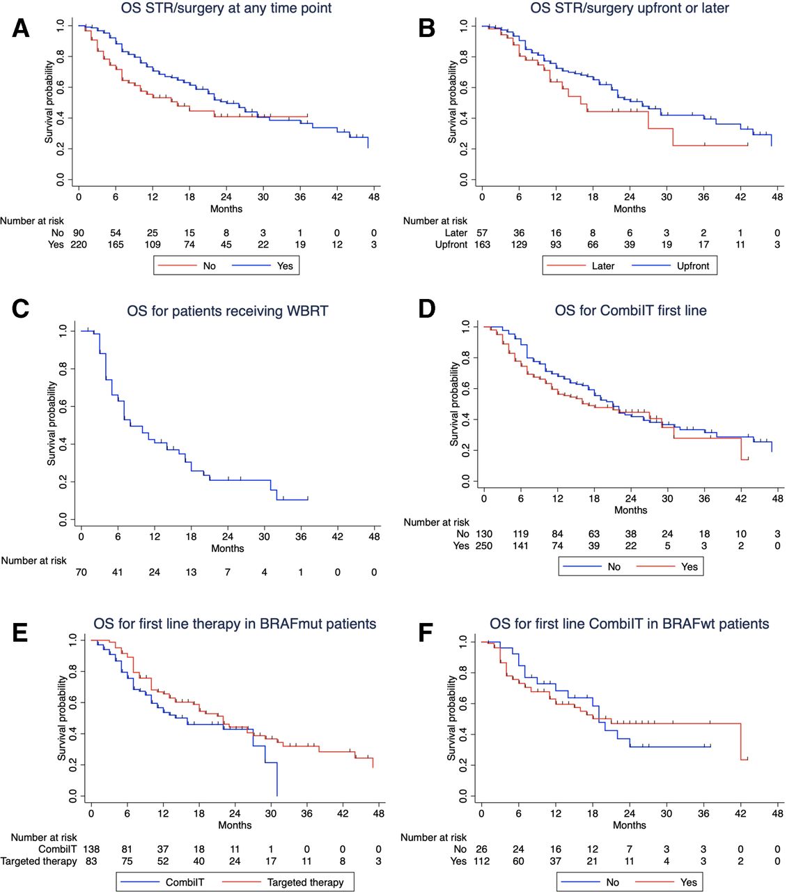
Local therapy (STR/surgery) also improved OS (table 3, figure 2A): patients who received local therapy (at any time point of the course of the disease) reached a mOS of 24 months compared with patients without local therapy with only 16 months (p=0.009; 95% CI: 19.6 to 28.4 and 7.6 to 24.4, respectively). There was no significant difference in terms of patients’ characteristics in these two groups, except for S100B level and presence of symptomatic MBM (online supplementary table S1). However, we need to acknowledge that information regarding the presence of symptomatic MBM was missing in approximately 50% of the patients.

Kaplan-Meier curves for overall survival (OS) according to the following factors: (A) local therapy (STR/surgery, stereotactic radiosurgery or surgery); (B) time of local therapy (before or after combined immunotherapy with nivolumab+ipilimumab); (C) for patients receiving whole brain radiotherapy (WBRT); (D) combined immunotherapy for melanoma brain metastasis (MBM) in first line or later; (E) first-line therapy in patients harboring a BRAF mutation and (F) combined immunotherapy first line or later in BRAF wild-type patients. Patients treated with WBRT were excluded from the analysis in figure 2A. Ten patients (4.5%) from the STR/surgery group (n=220) received only surgery. In the Kaplan-Meier analysis in figure 2B, four patients receiving STR/surgery before combined immunotherapy and two patients receiving STR/surgery after combined immunotherapy were treated with the two techniques in an interval of 2 weeks.
When analyzing the time point of local therapy (ie, before or after NIVO+IPI), we found no significant difference in terms of patients’ characteristics (online supplementary table S2) and the mOS was similar in the two subgroups (figure 2B; p=0.110). However, there seems to be a trend for a benefit of STR/surgery upfront (mOS=26 months vs 16 months; 95% CI: 21.1 to 30.9; 10.8 to 21.2, respectively). Patients who received WBRT had a mOS of 8 months (table 3, figure 2C; 95% CI: 4.9 to 11.0) and were analyzed separately.
No OS difference was observed for patients receiving first-line NIVO+IPI compared with those that received combined immunotherapy later (figure 2D; p=0.119). When looking at the patients’ characteristics from these two groups, there was a significant difference between them regarding age, BRAFV600 mutation status, number of MBM and treatment with local therapy (STR/surgery) at the time of starting NIVO+IPI (table 1). These differences might contribute for similar OS outcomes regardless of therapy line.
In the subgroup of patients with BRAFV600 mutation (242 patients), 83 received first-line treatment with BRAF/MEK inhibitors, 138 received first-line NIVO+IPI and all received combined immunotherapy for MBM in the course of the disease. There was no OS difference when comparing first-line targeted therapy with first-line combined immunotherapy (figure 2E; p=0.085). The line of treatment for combined immunotherapy (first-line or not first-line) had no effect on survival outcome in patients with BRAF wild-type melanoma (figure 2F; p=0.996).
Regarding presence of symptomatic MBM, information was available for only 193 patients (online supplementary figure S2D), but there is a trend benefiting patients with asymptomatic MBM (p=0.065). However, if we consider only the patients that received first-line NIVO+IPI for MBM (n=137), the difference in OS between symptomatic and asymptomatic MBM is not significant (p=0.084; data not shown).
Safety
In the present cohort, a total of 236 (62%) patients were reported to have at least one immune-related adverse events (irAEs). In 142 (37%) patients, no irAEs were documented and there was no available information in 2 (1%) patients.
We found no difference (Pearson’s χ2 test) in terms of onset of irAEs in patients with 1–3 MBM compared with patients with >3 MBM (p=0.069). Regarding the onset of irAEs in patients who received STR/surgery versus those who did not receive STR/surgery, there was also no significant difference between the two groups (p=0.657). Finally, when analyzing the relation between receiving STR/surgery or not, and the interruption of therapy due to irAEs, we again found no significant difference between the two groups (p=0.913).
Discussion
The present study shows that combined immunotherapy with NIVO+IPI can result in improved survival of patients with MBM, comparable to results in other stage IV patients. This is particularly true if intracranial CR, PR or SD has been achieved. The type of intracranial response is a strong predictor for OS. In our cohort, the 2-year OS rates of patients with SD, PR and CR ranged from 63% to 86%, whereas patients with PD had a 2-year OS rate of only 20% (table 3). Similar favorable results have been reported in the ABC trial, a randomized phase II study of nivolumab or NIVO+IPI in patients with MBM.16 The 3-year intracranial PFS was above 90% for patients with asymptomatic, treatment-naïve MBM achieving an intracranial CR, and above 50% for patients with PR. We have no explanation why in our cohort patients with SD did better than patients with PR.
In our study, the 1-year and 2-year OS rate were 69% and 41%, respectively, in line with previous reports.9 10 In the already mentioned ABC trial, patients who received combined immunotherapy had a 1-year and 2-year OS rate of 63%16 and in the Checkmate-204 trial the reported 1-year OS rate was even higher (81.5%).10 The survival rates in these trials are higher than those reported in our cohort. Compared with the ABC trial and the Checkmate-204 trial, which included patients with asymptomatic MBM and treatment-naïve BRAF wild-type patients, 31% (60/193) of the patients in our trial had symptomatic MBM and 20% of the BRAF wild-type patients were pretreated. In the Checkmate-204 trial, 17% of the patients had received previous systemic therapy for MBM and 52% had only one MBM compared with 34% pretreated patients and 53% patients with more than three MBM in our cohort.
Two studies evaluating pembrolizumab in patients with MBM also reported similar outcomes.17 18 The first study evaluated treatment with pembrolizumab monotherapy in 23 patients with one or more asymptomatic and untreated MBM. With a longer follow-up of 38 months, the mOS time was 17 months (95% CI: 10 months to not reached) and the 2-year OS was 48%. These are in line with our results for patients who did not receive STR/surgery for whom the mOS was 16 months (95% CI: 7.6 to 24.4) and the 2-year OS rate was 41%. However, in this trial, only asymptomatic patients were included and 87% had <3 MBM, a population with potentially better outcome that the one included in our report. In the second study, Anderson et al reported the results of the combination from pembrolizumab and radiation therapy in 21 patients with MBM. Despite the low number of patients included, the percentage of lesions that had a CR (>30%), was higher than previously reported with systemic therapy or STR alone.
The combination of immunotherapy and local therapy with stereotactic irradiation or surgery improved patients’ survival compared with patients who only received NIVO+IPI. This benefit might be related to a synergic effect between radiotherapy and immunotherapy that has been demonstrated both in preclinical and clinical studies.19–23 The combination of radiation and immune checkpoint inhibitors seems to be effective both in the irradiated and non-irradiated lesions, and this effect might be associated with the activation of cytotoxic T-cells and reduction of myeloid-derived suppressor cells.18 24 25
The benefit of combining local and systemic therapy in MBM has been previously shown by our group and others, with mOS that range from 14 to 25 months and 1-year OS rates between 58% and 78% in the groups that received local and systemic therapy, clearly superior to the outcomes of patients receiving only systemic therapy (mOS between 6 and 13 months and 1-year OS rates ranging from 34% to 53%).14 15 26–33
In our study, the time point at which the patients received local therapy did not seem to play a significant role in OS: local therapy performed upfront or after initiation of NIVO+IPI resulted in similar OS rates, with a trend benefiting local therapy upfront (mOS 26 months vs 16 months). Different retrospective studies have also addressed this question, and, similar to our cohort, upfront local therapy seems to have better outcomes (mOS of 11–23 months in the group receiving local therapy upfront and 3–9 months in patients receiving local therapy after systemic therapy).34 35
There is still an ongoing debate whether some patients might be better served with systemic therapy alone, as we see very positive outcomes.9–11 36 Not applying local therapy reduces local complications, potential cognitive impairment and might be particularly adequate for patients with a low number of asymptomatic MBM. This question along with the best sequence regarding local therapy is being addressed in ongoing clinical trials, and in the future, we might be better equipped to decide which patients to treat with the different modalities.37 38
In this study, there was a high proportion of patients with BRAFV600-mutated melanoma (63%), but similar to other publications where this subgroup represents between 52% and 65% of the patients.14 15 26 28 Previously, it has been postulated that even in patients with BRAFV600-mutated MBM, first-line systemic treatment should consist of combined immunotherapy. Our analysis showed that there was no difference in OS of patients receiving first-line NIVO+IPI or first-line targeted therapy followed by combined immunotherapy (p=0.085). The two subgroups did not differ significantly (online supplementary table S4), except for the number of MBM, where a higher proportion of patients with >3 MBM received first-line targeted therapy (p=0.002). Our results in this subgroup need to be interpreted with caution since we have not included patients with BRAFV600 mutation who only received targeted therapy.
In the multivariate Cox regression analysis, we identified LDH, S-100B, ECOG-PS and number of MBM as independent prognostic factors. These prognostic factors have already been described in previous analyses,8 14 39–41 but to the best of our knowledge, S100B has only been described as independent prognostic factor for checkpoint inhibitor immunotherapy in one monocentric study.42 It is interesting, however, that both tumor markers, LDH and S100B, remained independent prognostic factors in the multivariate analysis, suggesting that these non-invasive and easy to determine blood parameters can and should be used early in the course of the disease to inform about patients’ prognosis.
Regarding the presence of symptomatic MBM, there was no OS differences between patients with and without symptoms (p=0.065), but a trend can be seem showing that patients with symptomatic MBM have worse prognosis that those who are asymptomatic (1-year OS rate 46% and 63%, respectively). In other prospective studies investigating similar cohorts, the OS rate ranged from 66% at 6 months43 to 31% at 12 months.16 Unfortunately, information regarding the presence of symptomatic MBM is missing in approximately 50% of the patients in our study, and therefore, definitive conclusions cannot be drawn from our data.
Strengths of this investigation are that data from 23 German-certified skin cancer centers with high standards for data quality were included. Three-hundred and eighty patients were analyzed which is thus far the largest published cohort of patients with MBM managed in a routine clinical setting. This high number of patients allowed us to perform subgroup analyses, with results of reasonable sensitivity. Furthermore, this study provides long-term follow-up data of patients with MBM covering a period of up to 18 months.
The study limitations are related to its retrospective design. Patients were included regardless of previous systemic and local therapies prior to the combined immunotherapy and thus some heterogeneity of the study population might have contributed to differences in survival outcomes observed in our cohort. The decision to offer local therapy or not was probably influenced by the number and size of MBM. Additionally, the maximum number of MBM considered to be treated individually by STR/surgery might vary between different centers. We have not evaluated intracranial toxicities. However, this aspect might have been considered when planning local therapy and targeted therapy in patients with BRAFV600-mutated melanoma, influencing the systemic therapy offered as well as the therapy sequence in this subgroup.
In conclusion, our study shows that treatment with NIVO+IPI, particularly in combination with STR/surgery improves survival of patients with MBM. Results presented herein also suggest that local therapy with STR/surgery either before or after starting combined immunotherapy might be advantageous to prolonging OS.
References
Footnotes
Twitter @TeresaSAmaral
Contributors Study concept: TA, TE, CG, LZ. Data collection: all authors. Data analysis: TA, TE, CG, LZ. Data interpretation: TA, RG, CB, ChP, TG, JH, FM, TE, CG, LZ. Writing: all authors. Final approval: all authors. Agreement to be accountable for all aspects of the work: all authors.
Funding The authors have not declared a specific grant for this research from any funding agency in the public, commercial or not-for-profit sectors.
Competing interests TA: reports personal fees and travel grants from BMS, grants, personal fees and travel grants from Novartis, personal fees from Pierre Fabre, grants from Neracare, grants from Sanofi, outside the submitted work. FK: reports personal fees from Amgen, personal fees from BMS, personal fees from MSD, grants and personal fees from Novartis, personal fees from Pierre Fabre, personal fees from Roche, personal fees from Sanofi, outside the submitted work. CL: reports personal fees from BMS, personal fees from MSD, personal fees from Merck, personal fees from Novartis, personal fees from Roche, personal fees from Pierre Fabre, personal fees from Sanofi, personal fees from Amgen, personal fees from Biontech, personal fees from Sun Pharma, other from Kiowa Kirin, outside the submitted work. KK: reports grants and personal fees from BMS, personal fees from MSD, during the conduct of the study; personal fees from Amgen, grants and personal fees from NeraCare, grants and personal fees from Novartis, personal fees from Philogen, grants and personal fees from Roche, grants and personal fees from Sanofi, outside the submitted work. PT: reports personal fees from BMS, Novartis, MSD, Pierre Fabre, CureVac and Roche, personal fees from BMS, Novartis, Pierre Fabre, Merck Serono, Sanofi and Roche, non-financial support from BMS, Pierre Fabre and Roche, outside the submitted work. AG: reports personal fees from BMS, personal fees from MSD, personal fees from Novartis, personal fees from Pierre Fabre, personal fees from Pfizer, personal fees from Roche, personal fees from Sanofi, outside the submitted work. RG: reports honoraria: Almirall Hermal, Amgen, Bristol-Myers Squibb (BMS), Incyte, Merck Serono, MSD, Novartis, Pierre Fabre, Pfizer, Roche, SUN; research funding: Amgen, Johnson & Johnson, MerckSerono, Novartis, Pfizer; travel and accommodations: BMS, Merck Serono, Pierre Fabre, Roche, SUN, outside the submitted work. SH: reports grants and personal fees from BMS, personal fees from MSD, during the conduct of the study; personal fees from Amgen, personal fees from Novartis, personal fees from Roche, personal fees from Sanofi, personal fees from Pierre Fabre, outside the submitted work. JU: is on the advisory board or has received honoraria and travel support from Amgen, BMS, GSK, LeoPharma, Merck Sharp & Dohme (MSD), Novartis, Pierre Fabre, Roche, Sanofi outside the submitted work. CB: has been investigator of clinical trials sponsored by Amgen, Array Pharma, BMS, ImmunoCore, MSD, Novartis, Regeneron and Roche; has received speaker’s and/or consultant’s fees by Amgen, BMS, ImmunoCore, Merck, MSD, Novartis, Pierre Fabre, Roche, Sanofi-Aventis and SunPharma, outside the submitted work. DR-S: reports personal fees from Novartis, personal fees from Roche, outside the submitted work. AK: reports advisory board honoraria from Novartis Pharma, Roche, travel grants from Amgen and BMS, personal fees from AbbVie and Medac Pharma, outside the submitted work. ChP: reports personal fees from BMS, MSD, Roche, Pierre Fabre, Novatris and SUNPharma for advisory roles during the conduct of the study. TG: reports receiving speakers and/or advisory board honoraria from BMS, Sanofi-Genzyme, MSD, Novartis Pharma, Roche, AbbVie, Almirall, Janssen, Lilly, Pfizer, Pierre Fabre, outside the submitted work. ClP: reports personal fees from BMS, personal fees from MSD, during the conduct of the study; personal fees from Amgen, personal fees from Merck Serono, personal fees from Novartis, personal fees from Roche, personal fees from Sanofi, personal fees from Pierre Fabre, outside the submitted work. DD: reports consulting and speaking fees and/or payment of travel expenses/participation fees: Amgen, BMS, MSD, Mylan, Novartis, Pierre Fabre, Roche, Sanofi, outside the submitted work. RH: served as consultant and/or has received speakers’ honoraria from Roche, BMS, MSD, Novartis and Pierre Fabre outside the submitted work. SE: reports advice and speakers’ honoraria from MSD, BMS, Pierre Fabre, Sanofi, Amgen, Novartis, LEO Pharm, ROCHE and Genzyme Corporation, outside the submitted work. JCH: reports personal fees and travel grants from BMS, MSD, Novartis, Roche, Pfizer, Pierre Fabre and Sanofi, grants for scientific projects from BMS, personal fees for participation in advisory boards from MSD and Pierre Fabre, outside the submitted work. FM: reports personal fees and non-financial support from Novartis, personal fees and non-financial support from Roche, personal fees from MSD, personal fees and non-financial support from BMS, personal fees and non-financial support from Pierre Fabre, outside the submitted work. TT: reports grants and personal fees from Novartis, grants and personal fees from Roche, outside the submitted work. TE: reports personal fees from Amgen, grants and personal fees from BMS, personal fees from MSD, grants and personal fees from Novartis, personal fees from Pierre Fabre, grants and personal fees from Roche, grants and personal fees from Sanofi, outside the submitted work. CG: reports grants and personal fees from BMS, personal fees from MSD, during the conduct of the study; personal fees from Amgen, grants and personal fees from NeraCare, grants and personal fees from Novartis, personal fees from Philogen, grants and personal fees from Roche, grants and personal fees from Sanofi, outside the submitted work. LZ: served as consultant and/or has received honoraria from Roche, BMS, MSD, Novartis, Pierre Fabre, Sanofi, and travel support from MSD, BMS, Amgen, Pierre Fabre, Sanofi and Novartis, outside the submitted work.
Patient consent for publication Not required.
Ethics approval and consent to participate The current study was submitted and approved by the Ethics commission of the Eberhard′s Karls University Tuebingen (approval number: 766/2018BO2).
Provenance and peer review Not commissioned; externally peer reviewed.
Data availability statement Data are available on reasonable request. The data that support the findings of this study are available but restrictions apply to the availability of these data, which were used according to the Ethics Commission vote and recommendations for the current study, and so are not publicly available.