Article Text

Abstract
Purpose Patients with cancer receiving tumor-reactive humanized monoclonal antibody (mAb) therapy can develop a human antihuman antibody (HAHA) response against the therapeutic mAb. We evaluated for HAHA in patients with neuroblastoma treated in a phase I study of humanized anti-GD2 mAb (immunoglobulin (Ig)G1 isotype), hu14.18K322A (NCT00743496). The pretreatment sera (collected prior to mAb treatment) from 9 of 38 patients contained antitherapeutic antibodies, even though they had no prior mAb exposure. We sought to characterize these pre-existing antitherapeutic antibodies (PATA).
Experimental design The PATA+ pretreatment samples were characterized via ELISA; clinical associations with PATA status were evaluated.
Results Pretreatment sera from eight of nine PATA+ patients also bound rituximab and demonstrated preferential ELISA reactivity against the Fc portions of hu14.18K322A and rituximab as compared with the Fab portions of these mAbs. These PATA+ sera also recognized dinutuximab (human IgG1 isotype) and mouse IgG2a isotype mAbs, but not a mouse IgG1 isotype or the fully human panitumumab (IgG2 isotype) mAb. Of the 38 treated patients, only 4 patients (all in the PATA+ cohort) demonstrated no disease progression for >2.5 years without receiving further therapy (p=0.002).
Conclusions This study demonstrates an association between clinical outcome and the presence of PATA against determinant(s) on the Fc component of the therapeutic mAb, suggesting that the PATA may be playing a role in augmenting mAb-based antitumor effects. Further analyses for the presence of PATA in a larger cohort of patients with relapsed neuroblastoma, analyses of their clinical correlates, identification of their immunological targets, and potential antitumor mechanisms are warranted.
- pre-existing antibodies
- mAb
- anti-GD2
- hu14.18K322A
- rituximab
- immunotherapy
- human antihuman antibody (HAHA)
- neuroblastoma
- pre-existing antitherapeutic antibodies (PATA)
This is an open access article distributed in accordance with the Creative Commons Attribution Non Commercial (CC BY-NC 4.0) license, which permits others to distribute, remix, adapt, build upon this work non-commercially, and license their derivative works on different terms, provided the original work is properly cited, appropriate credit is given, any changes made indicated, and the use is non-commercial. See http://creativecommons.org/licenses/by-nc/4.0/.
Statistics from Altmetric.com
- pre-existing antibodies
- mAb
- anti-GD2
- hu14.18K322A
- rituximab
- immunotherapy
- human antihuman antibody (HAHA)
- neuroblastoma
- pre-existing antitherapeutic antibodies (PATA)
Background
Tumor-reactive monoclonal antibody (mAb) therapy is a targeted approach to cancer treatment. An increasing number of these mAbs have been clinically approved.1 However, in some patients, treatment with mAbs generates antibody responses against the therapeutic mAb.
With the use of murine, chimeric, or humanized/human mAbs, patients can generate human antimouse antibody, human antichimeric antibody (HACA), or human antihuman antibody (HAHA) responses against the therapeutic mAb.2–4 These antitherapeutic antibodies may potentially “neutralize” the therapeutic mAb via increased clearance and/or interference with the tumor-binding of the mAb. To minimize mAb immunogenicity, humanzed and fully human mAbs are now the main mAb class in clinical trials.5
A source of immunogenicity on humanized/human mAbs, distinct from the idiotype, is the antibody allotypes, the heritable allogeneic polymorphisms in the antibody constant regions.6 Most immunoglobulin (Ig)G1 mAbs (chimeric or fully human) in common clinical use express at least 1 of 4 well-described IgG1 allotypes (GM1, GM2, GM3, or GM17), and all express a common kappa light chain allotype (KM1, KM1,2, or KM3).6 In vivo exposure to an Ig with a non-self-allotype can induce antibodies against the foreign allotype.6
Dinutuximab (a chimeric anti-GD2 mAb, ch14.18), given together with interleukin 2 (IL2) and granulocyte-macrophage colony stimulating factor (GM-CSF), is the standard of care treatment for children with high-risk neuroblastoma.7 8 The SJGD2 phase I clinical trial conducted at St Jude Children’s Research Hospital (SJCRH) sought to test the efficacy of a variant anti-GD2 mAb, designated hu14.18K322A, for patients with recurrent/refractory neuroblastoma. While screening for HAHA in the phase I clinical trial of the hu14.18K322A,9 10 we found that 9 of 38 patients had antibodies against our therapeutic mAb (hu14.18K322A) prior to receiving any treatment, and thus demonstrated pretreatment antitherapeutic antibodies (PATA) recognizing the hu14.18K322A mAb. Here we will show that these PATA recognized a determinant(s) shared among other mAbs, distinct from the 14.18 idiotype. Furthermore, PATA+ patients showed improved outcome compared with PATA− patients after hu14.18K322A treatment.
Methods
Patients
Clinical and demographic data for all SJGD2 study patients have been published.9 11 Serum samples were sent to the University of Wisconsin (UW) for institutional review board (IRB)-approved immunological analyses.
HAHA ELISA
Our standard ELISA system “bridging” HAHA assay has been well described.2 3 12 13 Detailed methodologies are shown in the online supplementary materials. Based on the distribution of low optical density (OD) values for pretreatment samples, we designated OD values <0.7 as negative and values >0.7 as positive.
Supplemental material
Modified HAHA ELISA for PATA specificity
To evaluate the reactivity of PATA+ sera to other mAbs, using our standard HAHA ELISA method, we altered the capture-mAb component (rituximab, rituximab Fab, rituximab Fc, hu14.18K322A, hu14.18K322A-Fab, hu14.18K322A-Fc, panitumumab, mouse 14G2a, and mouse IgG1 or IgG2a) used to coat the plate. We verified these reagents were plate-bound using anti-IgG (Fc specific), antimouse IgG, or antikappa light chain antibodies. The detection agent used was biotinylated hu14.18-IL2 (hu14.18-mAb fused to IL2).14 By using this detection reagent, we could verify whether the PATA+ sample recognized a determinant shared between hu14.18-IL2 and the “test” (plate-bound) mAb by both biotin detection and by measuring captured IL-2.
ELISA assays to detect human IgM HAHA
Using our standard ELISA protocol (detailed in the online supplementary materials), ELISA plates coated with hu14.18K322A, followed by sample incubation, were tested for IgM binding.
Binding inhibition assay
Our previously described binding inhibition assay was used to quantify the ability of human serum samples to inhibit the binding of anti-GD2 mAb to its binding partner (1A7 anti-idiotypic mAb).4 In brief, patient serum mixed with 12.5 ng of hu14.18K322A was used in an ELISA system to quantify detectible hu14.18K322A.4
Antibody fragmentation
Fab/Fc fragments were generated from intact rituximab and hu14.18K322A mAbs using Pierce Fab Preparation Kit (catalogue #44985) per manufacturer’s instructions. Sample purity was verified via gel electrophoresis followed by Coomassie staining. Fab fragment binding to cell surface targets CD20 or GD2 was verified via flow cytometry.
Reagents
All reagents used are listed in the online supplementary materials.
Statistical analyses
The Van der Waerden test was used to compare whether the hu14.18K322A concentration peak level for PATA+ patients was different from the peak level for PATA− patients. Fisher exact tests were used to assess associations between PATA status and clinical variables. Two-tailed t-tests were used to compare clinical/laboratory parameters between the PATA+/PATA− groups. One-way analysis of variance testing, using Bonferroni adjustment for multiple comparisons, was used to compare ELISA tests of sera between PATA+/PATA− groups. Statistical analyses were performed using SAS V.9.4 and GraphPad Prism. Tukey’s multiple comparison test was used to compare PATA reactivity against intact mAb and mAb components to obtain adjusted p values .
Results
PATA was detected in the pretreatment sera from 9 of 38 evaluable patients from the SJGD2 phase I study of hu14.18K322A (figure 1A). These nine PATA+ patients had no prior exposure to hu14.18K322A or any other mAb. The PATA reactivity was due to antibody reactivity against hu14.18K322A, as indicated by the IgM antibody found in patient serum (figure 1B), and this antitherapeutic reactivity remained detectable throughout the 1-month treatment course (figure 1C). This stable reactivity pattern for these nine PATA+ patients is distinct from the pattern for the PATA−, HAHA+ patients, who had HAHA detected in their serum after treatment began (figure 1D), or those patients that remained PATA− and HAHA− during therapy (figure 1D).
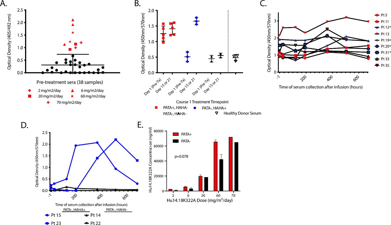
PATA against hu14.18K322A were detected in 9 of 38 patients. (A) OD values are shown from the HAHA assay for reactivity to hu14.18K322A for pretreatment sera for 38 evaluable patients. Twenty-nine samples (black circles) fell within the 95% CI of <0.7 OD (PATA−). Nine patients’ samples (non-circles) show reactivity >0.7 OD (PATA+). PATA− patients were found at every hu14.18K322A dose. PATA+ patients are classified by the dose of hu14.18K322A received (mg/m2/day): 2 (◆), 6 (▲), 20 (☐), 60 (✭), and 70 (+). (B) OD values for the ELISA assay to detect IgM binding to hu14.18K322A are shown for sera from 10 patients enrolled on the trial and three control serum samples obtained from 2 healthy donors. The OD readings of the patients are shown for pretreatment [day 1 (pre-Tx)] and post-treatment (day 15 or 21) sera. Five patients are PATA+, HAHA− (in red); three patients are PATA− and become HAHA+ (PATA−, HAHA+ in blue); two patients are PATA− and remain HAHA− (PATA−, HAHA− in black). (C) OD values for the HAHA assay for all nine PATA+ patients for course 1 serum time points (1-hour pretreatment, and 24, 120, 192, 360, 504, and 672 hours post-treatment). Patients 12, 19, 20, and 31 are the four patients who remained progression-free at 2.5 years (blue symbols and “*”). (D) OD values for the HAHA ELISA assay for four patients representative of two scenarios; patients 15 and 23 (in blue) were PATA− and became HAHA+ (at 192 or 360 hours); patients 14 and 22 (in black) remained PATA− and HAHA− and all their values are near 0 and appear superimposed. (E) For each of the hu14.18K322A treatment doses (doses of 2, 6, 20, 60, and 70 mg/m2/day) where PATA+ sera were observed in patients, the peak serum concentration of hu14.18K322A during course 1 for each of the PATA+ patients (n=9) and PATA− patients (n=17) were compared with dose as a covariate. At each hu14.18K322A treatment dose, while PATA+ patients had a higher peak serum concentration of hu14.18K322A level, there were no statistically significant differences in the peak serum concentration levels between PATA+ and PATA− patients at the different treatment dose levels (p=0.178). Note, this calculation excludes patients receiving 4, 10, 40, or 50 mg/m2 hu14.18K322A as none of the 12 patients treated at those doses were PATA+, and thus includes 9 PATA+ patients and 17 PATA− patients. HAHA, human antihuman antibody; Ig, immunoglobulin; mAbs, monoclonal antibodies; OD, optical density; PATA, pre-existing antitherapeutic antibodies.
In prior anti-GD2 clinical trials, we noted a decrease in peak serum levels of anti-GD2 mAb in treatment courses after the detection of the HACA or HAHA reactivity.4 In contrast to our previous observations, the majority of PATA+ patients had peak anti-GD2 mAb concentrations that were higher than that detected for PATA− patients treated at comparable hu14.18K322A doses (p=0.078, figure 1E). Although not statistically significant, consistent with figure 1E, PATA+ patients had increased levels for both area under the curve and half-life, commensurate with decreased clearance-levels of Hu14.18K322A, compared with PATA− patients (online supplementary figure 1). Thus, unlike HACA/HAHA, acquired after mAb exposure, PATA do not neutralize detectible serum levels of the therapeutic mAb.
In a binding inhibition assay, PATA+ serum samples do not inhibit hu14.18K322A from binding to its binding partner (1A7 mAb), thus PATA+ samples show no anti-idiotypic activity (table 1). In contrast, serum samples from a PATA− patient who developed HAHA after hu14.18K322A exposure and a pool of serum samples from patients who developed antitherapeutic antibody after exposure to other 14.18-based anti-GD2 mAbs4 15 show strong inhibition of hu14.18K322A binding to the 1A7 mAb, demonstrating induction of anti-idiotypic antibody (table 1).
Optical density in the HAHA assay and serological interference with the detection of hu14.18K322A
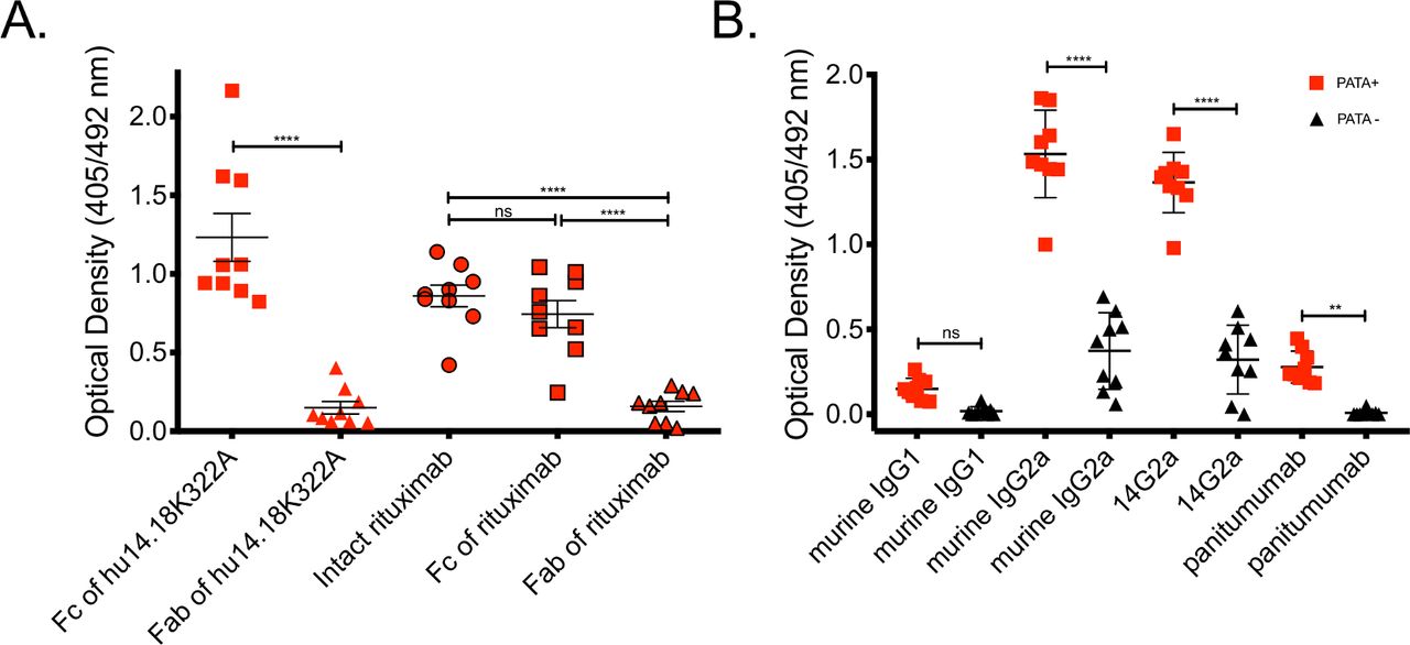
All PATA+ patients had PATA reactivity directed against the Fc component of hu14.18K322A (figure 2A), but not the Fab component. Serum from these PATA+ patients also recognized the Fc end of another mAb, rituximab. Rituximab (human anti-CD20) has the GM1,17 allotype, which is a different allotype than hu14.18K322A (GM3 allotype; online supplementary figure 2).6 Sera from eight of nine PATA+ patients bound to intact rituximab and the Fc fragment, but not the Fab fragment, of rituximab (figure 2A).
The antigenic target(s) of PATA are predominantly in the Fc region. A modified bridge ELISA was used to evaluate the reactivity of PATA+ sera to mAbs and their components. Only small serum volumes were obtained from each pediatric patient at each time point, thus insufficient pretreatment serum was available from some PATA+ patients for these assays. Reactivity against the Fab fragment of hu14.18K322A was observed using a serum sample previously identified as positive for anti-idiotype reactivity to hu14.18K322A, confirming that this ELISA system is capable of identifying anti-Fab reactive sera when present (data not shown). As the detection of PATA reactivity was stable between the pretreatment sample and the first post-treatment sample obtained 24 hours after starting treatment (figure 1C), and long before the acquisition of a new HAHA after exposure to hu14.18K322A (figure 1D), we used serum from the 24-hour post-treatment time point for the assays shown here. (A) OD readings from the nine PATA+ patients to the Fc portion of hu14.18K322A mAb, and the Fab portion of hu14.18K322A (non-outlined red symbols) and OD readings from each of nine PATA+ patients to intact rituximab, rituximab Fc, and rituximab Fab (black-outlined red symbols). In each column, the mean ±1 SEM is shown. ns (p=0.45), and ****(p<0.0001). (B) Sera from PATA+ patients recognize determinant(s) on murine IgG2a isotype antibodies and the fully murine anti-GD2 mAb 14G2a but show background reactivity to murine IgG1 mAb and panitumumab. An ELISA bridge assay was performed for all nine PATA+ patients (red squares) and for sera from nine representative PATA− patients (black triangles), assessing for the ability of the patients’ serum samples to bridge biotinylated hu14.18IL2 to various antibodies bound to the plate. ns (p=0.33), **(p<0.01), and ****(p<0.0001). IgG, immunoglobulin G; mAbs, monoclonal antibodies; OD, optical density; PATA, pre-existing antitherapeutic antibodies.
Figure 2B demonstrates that all nine PATA+ patients had significantly higher reactivity than the PATA− patients against a mouse IgG2a isotype control mAb and 14G2a (anti-GD2 mAb; mouse IgG2a Fc region). In contrast, the reactivity of sera from all PATA+ patients was not >0.7 OD cut-off against mouse IgG1 mAb isotype control or against panitumumab (anti-EGFR mAb; human IgG2 Fc region); there was no significant difference between PATA+ versus PATA− patients to recognize mouse IgG1 mAb, but there was a slight elevation in reactivity of PATA+ to panitumumab versus to PATA− patients.
Of the 38 patients enrolled in the phase I study, only 4 remained progression-free at 2.5 years after starting treatment (table 2). All 4 of these patients are in the PATA+ group of 9 patients; 0 of 29 PATA− patients were non-progressors (p=0.002; table 2). As prior studies of anti-GD2-based mAb agents suggest more likely benefit in the absence of bulky disease,16 the disease burden was evaluated qualitatively (online supplementary tables 1 and 2). The distribution of patients with high disease burden (classes 3 and 4) versus low disease burden (classes 1 and 2) was similar in PATA+ patients (5 with high burden and 4 with low burden) and PATA− patients (18 with low burden and 11 with high burden) (online supplementary tables 1 and 2). At 2.5 years after starting treatment, only the 4 PATA+ patients with low disease burden (all were class 2) had no disease progression without further therapy; all 34 other patients (including the 5 PATA+ patients with class 4 high disease burden and the 29 PATA− patients) showed progression (p<0.001; table 2).
Clinical characteristics of patients with detection of PATA and absence of disease progression
Discussion
To our knowledge, this is the first study describing an association of the presence of PATA with the improved clinical outcome of a tumor-reactive mAb trial. Although the specific epitope(s) are yet to be determined, we can rule out certain well-defined sources of mAb immunogenicity. PATA reactivity in this study cannot be accounted for by focusing only on the hu14.18K322A point mutation, as we detect PATA directed against rituximab, which does not contain this mutation. PATA is not directed against the 14.18 idiotype, as PATA did not diminish hu14.18K322A binding to 1A7 (table 1).
Human IgG1-based mAbs have well-characterized, serologically defined allotypes (four on the heavy chain and three on the kappa light chain) (online supplementary figure 2).6 Most IgG1 mAbs in clinical practice express a kappa light chain with a common allotype (KM3). Of the PATA+ patients, six of nine had evaluable DNA for genotyping; all six were found to have the KM3 allele (data not shown); they would be unlikely to make an antibody to KM3 as it would not be foreign. Further, the PATA reactivity binds primarily to the Fc region, rather than Fab, of both hu14.18K322A and rituximab (figure 2). Among the allotypes on the Fc fragment of IgG1, only one allotype (GM1) is expressed on rituximab (online supplementary figure 2). Of the six PATA+ patients (genotyped by pyrosequencing for GM allotypes), two of them were positive for GM1 (data not shown). GM1, GM2, and GM3 are not expressed on the Fc region of hu14.18K322A. Therefore, if sera from PATA+ patients recognize a region that is expressed on both hu14.18K322A and rituximab, then it must be a different allotype from the well-described allotypes found on human IgG1 (online supplementary figure 2).
None of the PATA+ patients strongly recognize the human IgG2 isotype, panitumumab, or a murine IgG1 isotype antibody. These data suggest that the epitope(s) recognized on hu14.18K322A by serum samples from PATA+ patients are not present on panitumumab or the murine IgG1 mAb tested. They also suggest that PATA reactivity against hu14.18K322A (produced in the rat myeloma cell line, YB2/0, with decreased fucosylation activity) is not directed against murine/rodent glycosylation patterning as both rituximab and panitumumab (recognized vs not recognized by PATA+) are produced in the CHO cell line, which has lower levels of glycosylation.17
Since identifying PATA in 9 of 38 patients in this trial, we considered why we had not observed PATA in past trials of anti-GD2 mAb using our bridge ELISA assays. In previous studies, our biotinylation kit recommended 20 molar excess of the biotinylation reagent. Recently, we optimized this to instead use 5–10 molar excess for our assays, resulting in adequate biotin-labeling while retaining mAb function/specificity. We confirmed that excess biotinylation interferes with allotype binding by reanalyzing samples from a COG phase III trial, ANBL0032,7 for which we had found that 0 of 126 patients evaluated demonstrated PATA in our previously used bridge ELISA assay. Using our optimized biotinylation protocol, we randomly selected 40 of these 126 patients and reassayed their pretreatment samples, and found that 3 of 40 patients (8%) demonstrated a PATA response (using the same 0.7 OD “cut-off”; data not shown). Thus, reduced biotinylation of hu14.18K322A allows for more sensitive detection of PATA than our prior assays, likely due to the excessive biotinylation causing blocking of the binding sites recognized by antiallotype mAbs.
Separately, we questioned whether the patient population we studied here was more likely to have PATA than other populations of cancer patients. In these patients with relapsed or refractory neuroblastoma, virtually all patients would have had combined, myelosuppressive chemotherapy, requiring multiple transfusions of blood products (particularly platelets) resulting in exposure to allogeneic blood products (plasma and Ig). We screened for PATA in a hu14.18K322A trial of newly diagnosed pediatric neuroblastoma patients, who had not yet had any therapeutic interventions or exposure to blood products (Trial NB2012 of SJCRH; NCT01857934).18 Of the first 32 patients enrolled in the study, none were PATA+ in their pretreatment samples; the incidence of PATA positivity in these two St Jude studies of hu14.18K322A (9/38 vs 0/32) was significantly different (p=0.003). Thus, prior exposure to allogeneic blood products may be inducing antibody responses to allogeneic epitopes shared between the therapeutic hu14.18K322A and the Igs of allogeneic blood product donors, that are seen as “foreign” by the immune systems of the PATA+ patients.
Prior studies of tumor-reactive mAbs have looked for associations of disease outcome and the detection of antitherapeutic antibodies and focused primarily on the importance of serum containing antibody to the therapeutic mAb that developed after known exposure to the therapeutic mAb. Correlations with favorable outcomes have been demonstrated,19–21 and others found outcome correlations with “anti-anti-idiotype” antibody formation.22 Yet, such correlations may be confounded as patients who form antitreatment antibodies may have stronger immune systems.23 Our findings suggest that antitherapeutic antibodies found in the serum of patients being treated with tumor-reactive mAbs may have different in vivo effects, depending on what component the antitherapeutic antibodies recognize on the tumor-reactive mAb. Our findings suggest that PATA may act in conjunction with the anti-GD2 mAb therapy, as PATA-associated benefit required hu14.18K322A treatment.
It will be helpful to define the molecular epitopes on therapeutic mAbs that are recognized by PATA. Sequencing the IgG constant regions for PATA+ versus PATA− patients may help test the hypothesis that PATA+ patients have subtle differences in their IgG Fc region compared with the Fc sequence from most therapeutic mAb, making the therapeutic mAb appear immunologically foreign to the PATA+ patients. If PATA+ patients have a better outcome when treated with tumor-reactive mAbs, then determining the mechanisms of PATA will be critical. The presence of PATA may increase the circulation time of the therapeutic mAb by extending its half-life, as figure 1E and online supplementary figure 1 suggest. Alternatively, PATA “cross-linking” of tumor-bound mAb on the tumor may enhance tumor killing via: (a) inducing tumor cell apoptosis24; (b) increasing mAb avidity to the antigen target; or (c) increasing the Fc density at the cell surface, thereby augmenting the antibody-dependent cell-mediated cytotoxicity or complement-dependent cytotoxicity potential (online supplementary figure 3).25
Conclusions
Several tumor-reactive mAbs show clear clinical efficacy. Identification of newer mAb targets on tumors will likely generate a clinical armamentarium of therapeutic mAbs, allowing for recognition of virtually any cancer type. Although some patients respond to tumor-reactive mAb-based treatments, many still do not. To increase the efficacy of mAb therapeutics, their mechanisms of action and interface with the immune system must be rigorously studied. The findings presented here support the possibility that tumor-reactive mAbs may interact with endogenous non-neutralizing serum antibodies in a manner augmenting antitumor effects. Further clarification of this phenomenon and the underlying mechanisms might provide leads for the development of more effective mAbs and mAb-based treatment regimens.
Acknowledgments
The authors thank Gwen Anthony, Lane Faughnan, Rupert Handgretinger, MD, Ray Barfield, MD, and St Jude clinical research nurses involved with this clinical trial and PATA protocol, Sharon H Moses and the UWCCC pharmacy for their helpful roles in the clinical trial and lab research enabling this analysis, critical discussion and provision of reagents, and the patients, families and clinicians who contributed to this research.
References
Footnotes
Correction notice Since the online publication of this article, the author names 'Jacquelyn A Hank' and 'Stephen D Gillies' were spelt incorrectly as 'Jacqueline A Hank' and 'Steven D Gillies'. This has now been corrected.
Contributors JLG: performed serological assays, interpreted the data, and was a major contributor to the writing of the manuscript. FN, VS, MWB, and MMM: were involved in clinical data collection, sample procurement, and were contributors in writing of the manuscript. JG, MM, and AMJ: performed serological assays, interpreted the data, and were contributors to the writing of the manuscript. JH, AE, FdB, and AH: involved in data interpretation and were contributors to the writing of the manuscript. LC and KK: performed statistical analysis and was a contributor to the writing of the manuscript. SDG: created the antibody studied, was involved in data interpretation, and was a contributor to the writing of the manuscript. JPP: was a major contributor to interpretation of the data regarding PATA and antibody allotypes and was a contributor to the writing of the manuscript. PMS: led the analyses assessing antibody levels in patients treated with the hu14.18K322A antibody, interpreted the results, and was a major contributor to the writing of the manuscript.
Funding This work was supported by the Alex’s Lemonade Stand Foundation, The St. Baldricks Foundation, The Howard Hughes Medical Institute, The MACC Fund, NIH Grants CA032685, CA87025, CA166105, CA197078, GM067386, UL1TR000427, 1TL1RR025013-01, Cancer Center Support Grants CA14520 and CA21765, The Stand Up To Cancer – St Baldrick’s Pediatric Cancer Dream Team Translational Research Grant (SU2C-AACR-DT1113), and the American Lebanese Syrian Associated Charities.
Competing interests Dr S. Gillies declares employment and ownership interests in Provenance Biopharmaceuticals.
Patient consent for publication Not required.
Ethics approval As approved in the initial clinical report (9), all patients signed approved consent forms. These correlative analyses reported in this manuscript were deemed by our institutional review board as exempt from human subjects research.
Provenance and peer review Not commissioned; externally peer reviewed.
Data availability statement Data are available upon reasonable request. The datasets used and/or analyzed during the current study are available from the corresponding author upon reasonable request.