Article Text

Abstract
Non-small cell lung cancer (NSCLC) can be associated with pulmonary cystic airspaces (pCAs). pCAs are radiologically classified into four types based on whether the nodule or mass extrudes the wall of the pCAs. In most cases, response evaluation of these lesions by Response Evaluation Criteria in Solid Tumors (RECIST) V.1.1 is challenging. Based on the observation of a case of morphological evolution of pCAs associated with NSCLC in a patient receiving immune checkpoint inhibitor (ICI), we reviewed retrospectively imaging scans of 92 consecutive advanced patients with NSCLC treated at our institution. Overall, three cases of pCAs associated with NSCLC obtained a remarkable change following ICI. Of note, these changes were not always seen in the context of a clear radiological objective response. The morphological changes observed may reflect a novel pattern of response to immunotherapy agents that clinicians should be aware of. This pattern of response, not reported before, warrants further investigation and, if confirmed, we believe that it should be considered in future version of immune RECIST.
- immunotherapy
- lung neoplasms
- programmed cell death 1 receptor
- case reports
This is an open access article distributed in accordance with the Creative Commons Attribution Non Commercial (CC BY-NC 4.0) license, which permits others to distribute, remix, adapt, build upon this work non-commercially, and license their derivative works on different terms, provided the original work is properly cited, appropriate credit is given, any changes made indicated, and the use is non-commercial. See http://creativecommons.org/licenses/by-nc/4.0/.
Statistics from Altmetric.com
Background
Non-small cell lung cancer (NSCLC) can be associated with pulmonary cystic airspaces (pCAs) in 1%–3.6% of cases.1 Radiological appearance of pCAs is classified in four types based on whether the nodule or mass extrudes the wall of the pCAs (type I), is confined within it (type II), forms a soft-tissue density extending along the pCAs wall (type III) or is intermixed within clusters of pCAs (type IV).2 In most cases, response evaluation of these lesions by Response Evaluation Criteria in Solid Tumors (RECIST) v1.13 is challenging.
Following the identification of a case of peculiar evolution of pCA lesions on immune checkpoint inhibitor (ICI) administration in a patient with NSCLC, we retrospectively evaluated CT scans of 92 consecutive advanced patients with NSCLC referred to our Medical Oncology Unit. All patients received ICI between 2015 and 2018. Radiological assessment, carried out by a radiologist with expertize in lung cancer imaging (MZ), was performed at baseline and following a period of at least 2 months, according to RECIST V.1.1.
Overall, we identified three cases (3.3%) of advanced patients with NSCLC with pCA lesions at baseline in which a relevant evolution in their morphology was observed following ICI therapy; of note, these changes were not always seen in the context of a clear radiological objective response.
Case presentation number 1
A 67-year-old heavy smoker woman was diagnosed with a Kirsten rat sarcoma virus oncogene (KRAS)-mutant anti-programmed cell death ligand protein 1 (PD-L1)-negative lung adenocarcinoma.
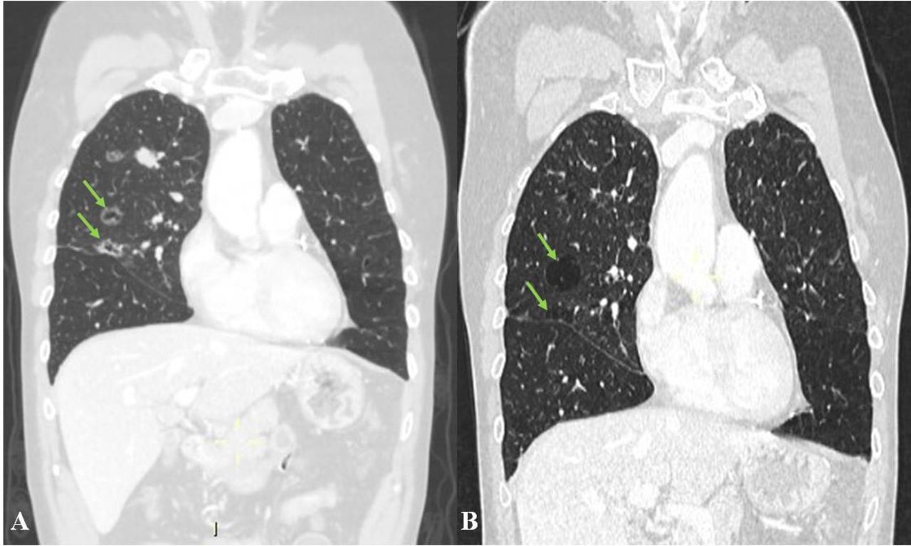
Baseline CT scan demonstrated a 50 mm solid mass in the left lower lobe with multiple bilateral lung metastases and type III pCA lesions (figure 1A). The patient received four courses of cisplatin-pemetrexed chemotherapy plus nivolumab, an anti-programmed death protein 1 (PD-1), followed by maintenance with pemetrexed and nivolumab.

Coronal CT images in lung window. Type III pulmonary cystic airspace lesions (green arrows) at baseline (A) and after (B) 17 months and 23 courses of chemotherapy and immunotherapy with nivolumab.
Disease assessment scan showed an objective response of the target solid lesion according to RECIST V.1.1 after about 17 months, while the pCA lesions evolved towards a larger-sized and thinner walled morphology (figure 1B). The changes observed in the pCA lesions, possibly reflecting reduction of cancer cells along their wall, might be considered as a radiological sign of treatment response. Maintenance chemotherapy is still ongoing without evidence of immune-related adverse event (irAE) or of disease progression, for a duration of response (DoR) of 20 months and progression-free survival (PFS) of 37 months.
Case presentation number 2
An 83-year-old former smoker woman received atezolizumab, PD-L1, as fourth-line treatment for her squamous PD-L1-positive (30%) NSCLC with lung and nodal metastases.
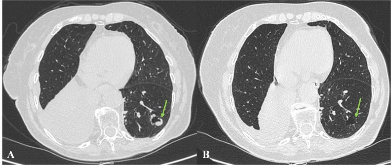
Baseline CT scan showed a 70 mm solid lesion in the right lower lobe. The examination also revealed a 21×16 mm thick-walled pCA lesion in the left lower lobe with mixed type I and II features (exophytic and endophytic abutting solid nodules), consistent with either lung metastases from NSCLC or concurrent second primary carcinomas (figure 2A).

Axial CT images in lung window. A left lower lobe pCA (mixed type I and II, green arrows) associated with squamous cell lung cancer, before (A) and after (B) 7 months and 10 courses of atezolizumab, showing resolution of both the solid component and the pCA lesion. pCA, pulmonary cystic airspace.
Radiological assessment after 5 cycles of atezolizumab demonstrated tumor response by RECIST V.1.1. After five further cycles, resolution of the solid component and of the wall thickening associated with the pCAs in the left lung was observed (figure 2B). The patient experienced hypothyroidism flare and was still alive and progression-free 11 months after atezolizumab starts, with an ongoing DoR of 9 months.
Case presentation number 3
An early 60s male smoker with stage IV KRAS-mutant, PD-L1-positive (75%) lung adenocarcinoma received pembrolizumab, another anti-PD-1 agent, as first-line treatment.
Baseline CT scan demonstrated a 42 mm solid mass in the left upper lobe together with a 45×34 mm subpleural pCA lesion with type II morphology (wall-thickened lesion with a solid nodule confined within the cystic airspace, figure 3A).

Axial CT images in lung window. A thin-walled pulmonary cystic airspace lesion (type II, green arrow) before (A) and after (B) 2 months and 2 courses of pembrolizumab treatment showing a stabilisation of the disease associated with remarkable increase of the solid component and filling of the airspace cavity.
The first radiological assessment showed a size decrease of the pCA lesion (maximum diameter 20 mm), with the airspace cavity filled by growth of the lesional solid component (figure 3B). This, together with decline of patient’s performance status, was more consistent with disease progression, despite disease was stable per RECIST V.1.1 on solid target lesion. The patient was next admitted and died 2 months after treatment starts (PFS: 2 months; overall survival (OS): 2 months). No irAE was observed.
Discussion pCAs with wall thickening or mural nodules are known signs of malignancy, and their identification can occur early in the diagnostic process of a pulmonary malignancy.1 4 The obstruction of small airways by tumor with induction of a check-valve mechanism and air trapping can induce the development of pCAs.5
Smoking history, KRAS mutations and adenocarcinoma histology are common in NSCLC associated with pCA lesions,6 but association with squamous histology has been reported as well.7
As opposite to our series describing lesions with a pre-existing cystic component, solid lesion evolution towards a cystic morphology has been observed as a response pattern on different types of treatment, especially vascular endothelial growth factor/receptor inhibitors.8–10
Despite the lack of pathology confirmation, the pCA lesions here described were likely malignant according to CT appearance and, based on their evolution, more probably represent metastases rather than secondary lung primary tumors.
Metastatic nature of pCA lesions was strengthened by the concordance of response with concomitant solid target lesions: in the first two cases, a complete vanishing of pCA lesions alongside a radiological response of solid pulmonary lesions was observed. By contrast, there was pCA lesion solid component growth in the third patient, whose disease was overall stable by RECIST V.1.1 but clinically progressed.
A previous report described a different case of lung tissue damage, defined as ‘bulla-like’ lesions, replacing lung metastases as an atypical response to nivolumab in a patient with NSCLC. The authors speculated on the role of T cell activation or autoimmune reaction to explain this atypical evolution of pulmonary metastases in permanent lung tissue injury.11
To our knowledge, this pCA-associated NSCLC evolution on ICI has never been reported and it differs from the previous report.11 Pattern of response can be characterized by reduction of wall thickening with increase in the air component, whereas pCAs can be filled by growth of the associated solid component with reduction of the airspace cavity in progressing ones.
Conclusions
RECIST V.1.1 evaluation of pCA lesions can be challenging and, as shown in this report, misleading in some cases. The morphological changes observed may reflect a novel pattern of response to immunotherapy agents that clinicians should be aware of.
Larger studies with systematic assessment of pCA lesions can be useful to further characterize radiological changes of this kind of lesions and their association with patterns of response on ICI and, if confirmed, this information should be incorporated in future version of immune RECIST.
Footnotes
Twitter @GLambertiMD
CP and GL contributed equally.
Contributors CP and MZ reviewed all imaging scans for each included patient. CP and GL reviewed medical history of case number 1 and prepared figure 1; FG and SS reviewed medical history of case number 2 and prepared figure 2; FS and AA reviewed medical history of case number 3 and prepared figure 3. CP, GL and FG wrote the manuscript with consultation with the other authors. FG and AA supervised the entire work. All authors contributed to the final version of the manuscript. All authors read and approved the final manuscript.
Funding The authors have not declared a specific grant for this research from any funding agency in the public, commercial or not-for-profit sectors.
Competing interests AA has received research grant support from BMS and Celgene; personal fees for serving in a consultant and/or advisory role for BMS, MSD and Boehringer; honoraria from Eli-Lilly and Pfizer.
Patient consent for publication Obtained.
Provenance and peer review Not commissioned; externally peer reviewed.