Article Text

Abstract
Background Talimogene laherparepvec (T-VEC) is a licensed therapy for use in melanoma patients of stage IIIB-IVM1a with injectable, unresectable metastatic lesions in Europe. Approval was based on the Oncovex Pivotal Trial in Melanoma study, which also included patients with distant metastases and demonstrated an overall response rate (ORR) of 40.5% and a complete response (CR) rate of 16.6%.
Objectives The aim of this study was to assess the outcome of melanoma patients treated with T-VEC in a real-life clinical setting.
Methods Based on data from 10 melanoma centers in Austria, Switzerland and southern Germany, we conducted a retrospective chart review, which included 88 patients (44 male, 44 female) with a median age of 72 years (range 36–95 years) treated with T-VEC during the period from May 2016 to January 2020.
Results 88 patients fulfilled the inclusion criteria for analysis. The ORR was 63.7%. 38 patients (43.2%) showed a CR, 18 (20.5%) had a partial response, 8 (9.1%) had stable disease and 24 (27.3%) patients had a progressive disease. The median treatment period was 19 weeks (range: 1–65), an average of 11 doses (range: 1–36) were applied. 39 (45.3%) patients developed adverse events, mostly mild, grade I (64.1%).
Conclusion This real-life cohort treatment with T-VEC showed a high ORR and a large number of durable CRs.
- oncolytic viruses
- immunotherapy
- melanoma
- oncolytic virotherapy
Data availability statement
All data relevant to the study are included in the article or uploaded as online supplementary information.
This is an open access article distributed in accordance with the Creative Commons Attribution Non Commercial (CC BY-NC 4.0) license, which permits others to distribute, remix, adapt, build upon this work non-commercially, and license their derivative works on different terms, provided the original work is properly cited, appropriate credit is given, any changes made indicated, and the use is non-commercial. See http://creativecommons.org/licenses/by-nc/4.0/.
Statistics from Altmetric.com
Introduction
Talimogene laherparepvec (T-VEC) is the first approved intralesional oncolytic therapy in the European Union, the USA and Australia for the treatment of unresectable stage IIIB, IIIC or IVM1a melanoma in Europe and up to IVM1c melanoma in the USA.1 2 It is injected directly into metastatic lesions.3 T-VEC is a genetically modified oncolytic herpes simplex virus type 1 (HSV-1).4 HSV-1 is modified through (1) the deletion of a neurovirulence gene (ICP34.5) and a immunogenicity gene (ICP47) and (2) the insertion of two gene copies encoded for human granulocyte macrophage colony-stimulating factor (GM-CSF).5 6 These modifications enhance tumor-selective replication, reduce virally mediated suppression of antigen presentation, and induce tumor-specific T-cell responses. Approval, in 2015, was based on the results of the phase III study Oncovex Pivotal Trial in Melanoma (OPTiM).1 7 Intralesional treatment with T-VEC, which was compared with subcutaneous application of human GM-CSF led to durable responses over at least 6 months, the primary endpoint of the OPTIM trial, in 25,2% of patients over only 1.2% in the GM-CSF arm. While the impact of T-VEC on overall survival (OS) did not show significance in the intention to treat population it did significantly improve OS in patients with stage IIIB, IIIC and IVM1a in a descriptive post hoc analysis.6 8 9
Well-implemented systemic therapies, like checkpoint inhibitors (CTLA-4, PD-1 Inhibitors)10–12 and targeted therapies (BRAF and MEK inhibitors) dramatically improved survival of patients with metastatic melanoma.13 However, large multicenter studies of checkpoint inhibitors and BRAF and MEK inhibitors did not pre-specify the subgroup of stage IIIB–IVM1a melanoma patients during recruitment.11 This subgroup was analyzed retrospectively with limited conclusions due to the small number of patients.11 14 15 Exactly this subgroup of patients with an initial low tumor burden might benefit from a treatment with T-VEC as an alternative treatment option to checkpoint inhibitors and targeted therapies, which in case of progression would still be available. The aim of this study was to assess the outcome of melanoma patients treated with T-VEC in a real-life clinical setting.
Material and methods
We performed a multi-institutional retrospective analysis of melanoma patients treated with T-VEC within the period of May 2016 and January 2020 (1370 days) (data cut-off).
Study site selection
Data were provided by 10 melanoma centers in Austria (AT), Switzerland (CH) and Germany (DE): Medical University of Vienna AT, University Hospital St. Poelten AT, Ordensklinikum Linz AT, Medical University of Graz AT, Landeskrankenhaus Klagenfurt AT, Krankenhaus der Elisabethinen AT, Krankenanstalt Rudolfstiftung, AT, University Hospital Zürich and Service d'Oncologie, CH, Center hospitalier Universitaire Vaudois, Lausanne, CH; Technical University of Munich, DE.
Study population
All participating centers were asked to provide data on all patients treated with T-VEC during the study period to avoid selection bias. Patients treated outside the approved indication and patients treated concurrently with other drugs were also included to provide a precise picture of real-life use of T-VEC. In total, 88 patients with a median age of 72 years (range 36–95 years), 44 male, 44 female, diagnosed with metastatic melanoma stage IIIB–IVM1d, based on the American Joint Committee on Cancer (AJCC V.8.0), were included in this retrospective chart review.
Data collection
Anonymized data were collected and entered in an electronic case report form. Patient data included demographics, melanoma history (primary melanoma diagnosis, tumor characteristics, anatomical region and mutation status), clinical characteristics (Eastern Cooperative Oncology Group (ECOG) Performance Status and comorbidities), laboratory parameters (lactate dehydrogenase (LDH), differential blood count, S100), the use of other melanoma therapies (type, duration) before, during, and after therapy with T-VEC, and finally the investigator assessed response rates, safety, and survival outcomes on T-VEC (table 1). Best overall response (BOR) was classified based on Investigator’s assessment as the best response achieved during the treatment with T-VEC. Due to the retrospective nature of this cohort investigator-assessed responses could be variably based on either measurement, count of metastases, clinical photographs or radiological assessments.
Patient data before and following the first dose of T-VEC
Statistical analysis
Descriptive statistics were provided for demographic, safety and efficacy analyzes. For nominally scaled variables we present absolute numbers and percentages. For metric variables mean, SD, median, minimum and maximum are provided. P values less than 0.05 are considered significant. Statistical analyzes were conducted using the statistic programs SPSS (V.26.0). Kaplan-Meier methods were used to estimate treatment persistence.
Results
Study population
Data were abstracted from 88 patients’ medical charts. The decision diagram is illustrated as a flow chart in figure 1. At the time of data abstraction, 6 patients (6.8%) had ongoing treatment and 82 patients (93.2%) had discontinued treatment. The reasons for discontinuation were as follows: (1) a complete response (CR) in 38 patients, (2) a stable disease (SD) in 8 patients, (3) a partial response (PR) in 18 patients, (4) a progressive disease (PD) in 24 patients and (3) adverse events (AEs) in 2 patients. Staging of disease was based on the AJCC criteria V.8.0: 9 patients (10.2%) were stage IIIB, 47 (53.4%) stage IIIC, 1 (1.1%) stage IIID, 18 (20.5%) stage IVM1a, 5 (5.7%) stage IVM1b, 4 (4.5%) stage IVM1c and 4 (4.5%) stage IVM1d. Demographics and baseline characteristics are presented in table 2. Similar numbers of patients were treated on an annual basis during the study period with a slight peak during 2018 (figure 2).

Flow chart of patients from centers in Austria, Switzerland and Germany included in the study. T-VEC, Talimogene laherparepvec.
Demographics and baseline characteristics
Primary tumor characteristics
The median primary tumor thickness Breslow Index within our patient cohort was 2.9 mm (range 0.35–40.00 mm). All patients underwent surgery on their primary melanoma with an adequate safety margin excision. In 74 patients (84.1%) a sentinel lymph node biopsy was performed, of which 38 (43.2%) were positive. Additionally, the mutation status information of melanoma metastases was collected (BRAF, NRAS, c-KIT). In 46 patients (52.3%) a mutation was detected: 31 patients (35.2%) had a BRAF mutation, 3 (3.4%) had a c-KIT and 12 (13.7%) had an NRAS mutation (table 2). The melanoma metastases were located as follows; trunk: 8 patients (9.1%), head: 12 (13.6%), lower extremities: 57 (64.8%), and upper extremities: 8 (9.1%) (table 2).
Clinical parameters and comorbidities
ECOG performance status (0–5) was ECOG 0 for 68 patients (77.3%), ECOG 1 for 17 patients (19.3%) and ECOG 3 for 3 patients (3.4%) (table 2). Comorbidities were as follows: arterial hypertension in 37 patients (42.0%), diabetes mellitus in 9 patients (10.2%), 15 patients (17.0%) had a history of a second malignancy, 1 patient (1.1%) had a history of organ transplantation, 3 (3.4%) had a chronic obstructive lung disease, 31 patients (36.0%) had a positive herpes simplex anamnesis.
Administration of T-VEC
T-VEC was administered into injectable metastatic melanoma lesions (cutaneous, subcutaneous and nodal tumors). The initial dose on day 1 was 106 plaque-forming units (PFU)/mL (up to 4 mL based on the lesion size). The second dose on day 21 was 108 PFU/mL (up to 4 mL based on the lesion size), the following cycles were applied every 14 days thereafter with 108 PFU/mL (up to 4 mL based on the lesion size). The median treatment period was 19.0 weeks (range: 1–65), an average of 11 doses (range: 1–36) were applied.
Efficacy outcomes
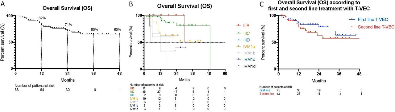
Eighty-eight patients fulfilled the criteria for analyzes. Investigator-assessed responses are shown in table 3. The overall response rate (ORR) was 63.7%. 38 patients (43.2%) showed a CR, 18 (20.5%) had a PR, 8 (9.1%) had an SD and 24 (27.3%) patients had a PD. The 45 patients (51.1%) treated with T-VEC as first line therapy showed better response rates, however differences where not statistically significant (p=0.185), compared with the remaining 43 patients (48.9%) treated with T-VEC as second line therapy (table 4). The BOR from patients treated with T-VEC as first-line therapy correlated significantly with longer progression free survival (PFS) (p=0.04), but did not correlate significantly with longer OS (p=0.199).
Investigator assessed best overall response.
Talimogene laherparepvec (T-VEC) as first-line therapy correlated with best overall response rates (complete response (CR), stable disease (SD), partial response (PR), progressive disease (PD))
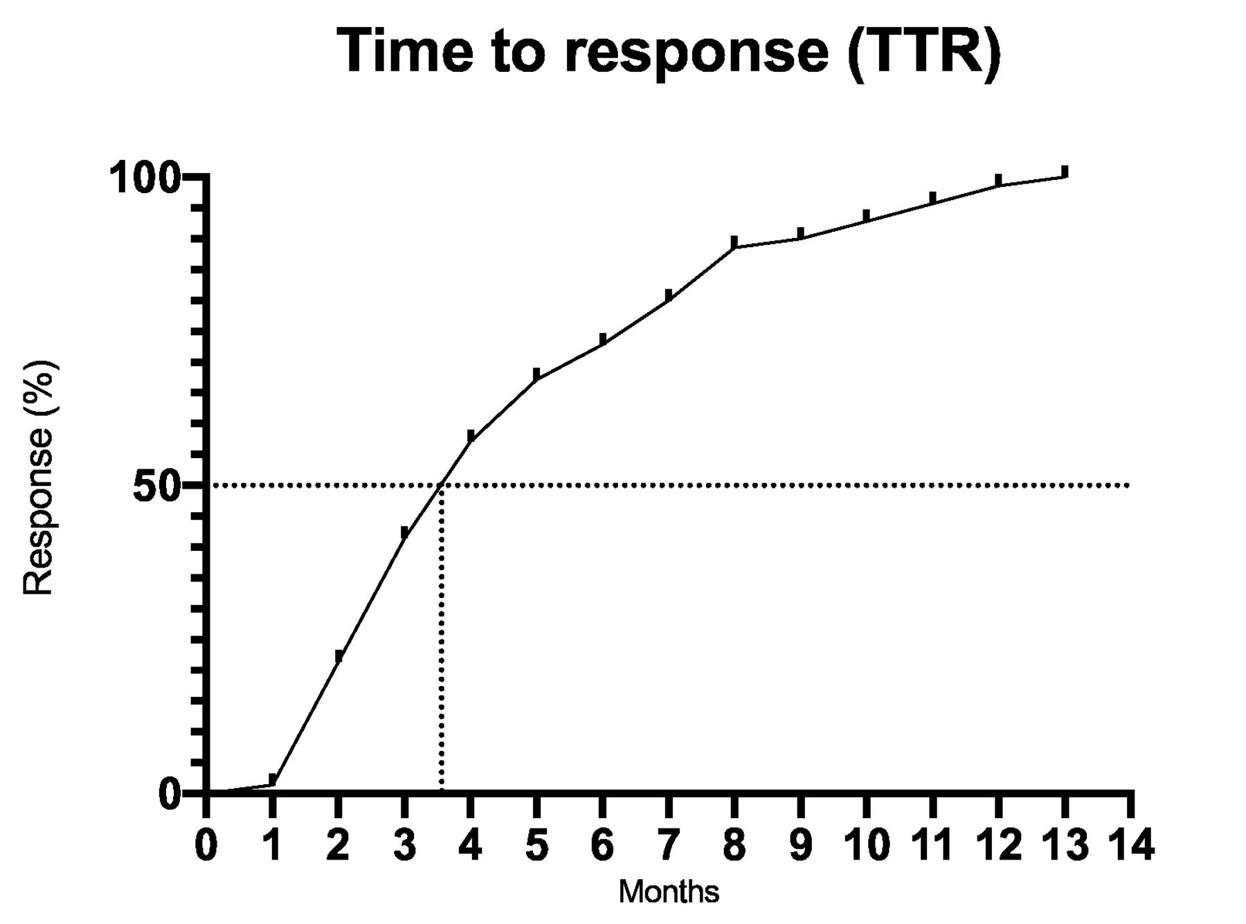
The median time to response was 124 days (range: 44–397) (figure 3). The median PFS was 9 months (95% CI 5.9 to 10.12) (figure 4A). The median OS was not reached (figure 5A). At the 1-year landmark 45% of all patients were without progression and 82% of patients were still alive (figures 4A, 5A). PFS (p=0.011) and OS (p=0.004) were significantly worse in patients with stage IVM1b to IVM1d melanoma (figures 4B, 5B). The 45 patients (51.1%) treated with T-VEC as first line therapy showed significant improved PFS (p=0.016) (figure 4C) and a trend toward improved OS (p=0.267) (figure 5C). However, OS differences were not statistically significant, compared with the remaining 43 patients (48.9%) treated with T-VEC as second line. The median follow-up period was 542 days (range: 14–1463 days). During the follow-up period 58 (65.9%) patients had a progression of disease: 32 (36.4%) patients progressed locoregional, 16 (18.2%) developed distant metastasis and 10 (11.4%) developed both, locoregional and distant metastases.

Time to response. Median time to response was 4 months=124 days (range: 44–397 days).

Kaplan-Maier analysis of PFS. One-year PFS was 45%, 2-year PFS was 35% and 3-year PFS was 28%. Median PFS was 9 months (95% CI 5.9 to 10.1) (A). Kaplan-Maier analysis of PFS according to disease stage (B). PFS according to first and second line treatment with Talimogene laherparepvec (T-VEC) (C). T-VEC, Talimogene laherparepvec.

Kaplan-Maier analysis of OS. One-year OS was 82%, 2-year OS was 71%, 3-year OS was 65% and 4-year OS was 65%. Median OS was not reached (A). Kaplan-Maier analysis of OS according to disease stage (B). OS according to first and second line treatment with tlimogene laherparepvec (T-VEC) (C). Kaplan-Maier analysis of PFS. One-year PFS was 45%, 2-year PFS was 35% and 3-year PFS was 28%. Median PFS was 9 months (95% CI 5.9 to 10.1) (A).

Talimogene laherparepvec (T-VEC) treatment over the years 2016–2020, in 10 melanoma centers in Austria, Switzerland and Germany. CPI, checkpoint inhibitors.
Laboratory parameters
Prior to therapy with T-VEC, the following laboratory parameters were collected: Lymphocytes (G/L), leucocytes (G/L), eosinophils (G/L), C reactive protein (CRP) (mg/dl), LDH and S100 (µg/L). Elevated S100 correlated with decreased PFS (p=0.0046). There was no significant association between eosinophils, lymphocytes, leucocytes, LDH and CRP and OS and PFS. Baseline laboratory parameters grouped in upper limits of normal and lower limits of normal based on BOR, namely CR, SD, PR, PD are shown in table 5.
Blood biomarkers: lymphocytes (G/L), leucocytes (G/L), eosinophils (G/L), C reactive protein (CRP) (mg/dL), lactate dehydrogenase (LDH) and S100 (ug/L) correlated with response rates
Use of other melanoma therapies before, during or after treatment with T-VEC
Tumor therapies used before, during or after T-VEC are shown in table 6. Forty-five patients (51.1%) received T-VEC as the sole melanoma therapy without prior antineoplastic treatment. Forty-three patients (48.9%) received therapy prior to treatment with T-VEC: 3 (3.4%) received radiotherapy, 12 (13.6%) PD-1 inhibitors, 10 (11.4%) Interferon, 5 (5.7%) BRAF and MEK inhibitors, 1 (1.1%) Imiquimod and 3 (3.4%) electrochemotherapy. A further nine patients (10.2%) received prior but unknown therapy.
Tumor therapies used before, during or after therapy with talimogene laherparepvec (T-VEC)
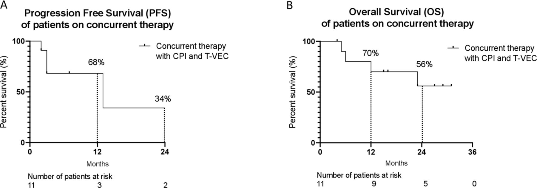
In addition to T-VEC, 11 patients (12.5%) received concurrent treatment: 10 patients (11.4%) PD-1 inhibitors and 1 patient (1.1%) a CTLA-4 inhibitor. These patients had advanced tumor stages: one patient stage IIIC, two patients stage IVM1a, two patients stage IVM1b, three patients stage IVM1c and three patients stage IVM1d. Eight of these 11 patients did receive T-VEC as an add on therapy on progression on a PD-1 inhibitor (5/8) or PD-1 based combination treatment (3/8). One patient received CTLA4 +T VEC on progression on a PD1 inhibitor and two patients received first line combinations of T-VEC with a PD-1 inhibitor. PFS and OS for patients on concurrent therapy are shown in figure 6A,B.
PFS of patients on concurrent therapy with checkpoint inhibitors (CPI) and talimogene laherparepvec (T-VEC). One-year PFS was 68%, 2-year PFS was 34%. The median PFS was 13 months (95% CI 10.0 to 15.3) (A). OS of patients on concurrent therapy with CPI and T-VEC. One-year OS was 70% and the 2- year OS was 56%. The median OS was not reached (B).
Thirty-three patients (37.5%) required therapy during the follow-up period. A detailed outline of follow-up therapies according to BOR and first or second line therapy with T-VEC is presented in table 7.
Follow-up therapy according to best overall response (BOR) (complete response (CR), stable disease (SD), Partial response (PR) and progressive disease (PD)) and first and second line therapy with talimogene laherparepvec (T-VEC)
Tolerability and safety
AEs were classified based on the common terminology criteria for AE V.5.0 on severity from grade 1 to 5. AEs were reported in 39 (44.3%) of the patients; 26 patients (29.5%) developed grade 1 AEs, 16 (18.2%) grade 2 AEs, 2 (2.3%) grade 3 AEs, 2 (2.3%) grade 4 AEs. No grade 5 AE occurred. Most common AEs were influenza like symptoms such as fever 21.6% (n=19), shivering 6.8% (n=6) and fatigue 8.1% (n=7). Herpetic lesions appeared in only one patient (1.1%) (table 8). Among the 11 patients that received concurrent treatment with T-VEC and checkpoint inhibitors, 5 (45.5%) developed AEs. Out of the remaining 77 patients which were treated only with T-VEC, 36 (46.8%) developed AEs.
AEs (adverse events): classified based on the CTCAE criteria V.5.0 on severity from grade 1 to 5
Discussion
There is an increasing amount of clinical data that supports the efficacy of T-VEC in treating metastatic melanoma.16–20 In our analysis, we provide data from an international cooperation across AT, CH and DE, countries that have similar access to novel melanoma treatments and follow comparable treatment standards in the management of metastatic melanoma. We provide detailed insights of patients treated with T-VEC in routine clinical practice.
Compared with 25.7 weeks in the OPTiM trial (which led to the approval of T-VEC), the median duration of T-VEC treatment was 19.0 weeks, whereas the ORR in our patients was 63.7%, vs 26.4% in the OPTiM trial.21 Our study included a population of patients with disease stages ranging from IIIB to IVM1d. 85.3% of the patients had stage IIIB–IVM1a and only a minority, 14.7%, had stage IVM1b–IVM1d. 51.1% of the patients in our cohort received T-VEC as first-line therapy. In the OPTiM study, only 55% of the patients were in stage IIIB-IVM1a and 47% of patients received T-VEC as first line therapy. Therefore, patient selection, also in relation to the European label for T-VEC, is the most likely explanation for the observed difference in ORR.
In other published real-life data analyzes, study populations with differing tumor stages were included. In the cosmus-1 trial, only 55.3% of patients had earlier stage IIIB-IVM1a metastatic melanoma, whereas other recent publications included only stage IIIB–IVM1a metastatic melanoma. In the cosmus-1 trial, 19.7% of the patients had a CR, however, the ORR was not evaluated.17–20 22 23 A multicenter retrospective German study, which included 27 patients with unresectable early stage IIIB–IVM1a melanoma treated with T-VEC, reported that 63% of the patient cohort received T-VEC as first-line therapy. The ORR was not evaluated.19 In a multicenter US study, conducted between 2015 and 2018, in which 42.5% of patients received T-VEC as first-line therapy the ORR was 56.4%.20 A single-site study of 26 T-VEC-treated patients from the Netherlands presented a similar safety profile and an ORR of 88.5%.24
While the majority of our patients were treated within the approved European indication, a minority of our patients with stage IVM1b–d received T-VEC as well. These were mostly patients with stable systemic disease, but locoregional progression, that received T-VEC as an add-on therapy. Responses have been observed in some of these patients. In our cohort, 11 patients (12.5%) received concurrent therapy with PD-1 or CTLA-4 checkpoint inhibitors. 22 patients (25.0%) received treatment with checkpoint inhibitors following progression on T-VEC. Due to the low number of patients requiring follow-up or concurrent treatment in our cohort we cannot assess if treatment with T-VEC did alter the response to a subsequent or concurrent systemic immunotherapy. The possibility that a local induction of an anti-tumor immune response could alter the response to systemic therapy with checkpoint inhibitors like PD-1 or CTLA-4 blocking antibodies has been addressed in early clinical trials and randomized studies to evaluate this possibility are currently ongoing in metastatic melanoma patients with injectable lesions.25–29
An analysis of the type of follow-up therapy according to BOR and first or second line treatment with T-VEC shows that patients receiving T-VEC as a first line therapy more often received a PD-1 inhibitor therapy compared with patients receiving T-VEC as a second-line therapy, which is in line with current recommendations to use PD-1 inhibitors as systemic first line treatment.30 From all the laboratory values collected at baseline only elevated S100 was associated with decreased PFS (p=0.046). The tolerability of T-VEC was similar to the OPTiM trial and other real-life studies with only 2 out of 88 patients stopping treatment due to AEs.9 17–20 22 23 The most common grade 1 and 2 AEs were fever 21.6% (n=19), chills 6.8% (n=6), fatigue 8.0% (n=7) and pain on the injection site 5.8% (n=5). There were 2 grade 3 AEs, namely cellulites on the injection site and colitis. One grade 3 AE colitis, one grade 4 cardiac AE and one grade 4 gastrointestinal AE occurred in patients who received simultaneous therapy with PD-1 inhibitors. Only one patient (1.1%) reported cold sores. T-VEC was well-tolerated, even though the study cohort represents an elderly population with a median age of 72 years and multiple comorbidities, with one patient even having a history of organ transplantation.31 In our cohort, no difference in the frequency of occurrence of AEs was observed between the patients who received T-VEC in combination with immunotherapy (45.5%) and patients who received T-VEC without immunotherapy (46.8%). While the documentation of AEs is usually less stringent outside of clinical trials, the recorded AEs are more likely to represent clinically significant events.
The era of checkpoint inhibitors and targeted therapies like BRAF and MEK inhibitors has led to dramatic improvements in OS and PFS for patients with metastatic melanoma and these drugs are the current standard therapies in the adjuvant as well as in the inoperable metastatic setting. In the latter, the 5-year OS rate for PD-1 inhibitor based therapies is between 39% and 52%,10 and for BRAF and MEK inhibitors up to 34%.13 However, more than 50% of patients do not respond to these and between 10% and 42% of patients, depending on the type of therapy have to stop these treatments because of AEs.32 Furthermore, despite the great effectivity of these drugs, the number of lines of therapies with a proven impact on OS is still limited for melanoma patients. Therefore, an additional treatment option that, as shown in our data collection, can add to the number of patients that achieve control of their disease is a clear advantage. We believe that T-VEC with its low toxicity profile is an ideal treatment option for selected patients with unresectable, but still limited cutaneous or subcutaneous metastases, especially for elderly patients or patients with multiple comorbidities. Checkpoint inhibitors and BRAF and MEK inhibitors would still be available to these patients in case of a potential recurrence of disease.
The strengths of our study include the size of the study population, the insights provided by a heterogeneous group of patients with different disease stages, clear and comprehensive information about clinical parameters, medical history and laboratory parameters. The main limitation of our study is its retrospective character which naturally limits the size, the depth and the availability of the data.
Conclusion
In our real-life cohort, treatment with T-VEC showed a high ORR and CR rate. Our findings support that T-VEC is a well-tolerated therapy that can be successfully used in patients with unresectable, locoregional and injectable metastatic melanoma. Elderly patients with multiple comorbidities who may not bear the risk of AEs from other systemic therapies and patients with a low tumor burden at the beginning of the treatment, might benefit specifically from T-VEC therapy.
Data availability statement
All data relevant to the study are included in the article or uploaded as online supplementary information.
Ethics statements
Patient consent for publication
Ethics approval
This study was registered at the Medical University of Vienna with the ethics committee number 18/40 2019.
References
Footnotes
Correction notice This article has been corrected since it first published. The provenance and peer review statement has been included.
Contributors Study design and supervision: CHO and JMR. Contributed in data collection: JMR, MK, LK, RS, JM, SL, VA, HK, FW, PK, CP, JK, OM, ER, CHA and CHO. Analyzed the data: JMR, RS and CHO. Wrote the paper: JMR, RS and CHO.
Funding The authors have not declared a specific grant for this research from any funding agency in the public, commercial or not-for-profit sectors.
Competing interests JMR received project funding by Amgen, Speakers bureau of Amgen and Bristol Myers Squibb and travel support from Bristol Myers Squibb, Pierre Fabre outside of the submitted work. JMR has intermittent project focused consultant or advisory relationships with Merck/Pfizer, Merck Sharp & Dohme, Amgen, Novartis, Bristol Myers and Squibb and Pierre Fabre and has received travel support from Ultrasun, L’ oreal, Merck Sharp & Dohme, Bristol Myers and Squibb and Pierre Fabre outside of the submitted work. PK has received honoraria for travel/congress support and consulting/advisory roles for Roche, Bristol Myers Squibb (BMS), Merck Sharp and Dome (MSD), Novartis, Amgen, Pierre Fabre and Sanofi Aventis unrelated to the submitted work. ER Honoraria, consulting or advisory role: Amgen, Bayer, Bristol Myers Squibb, MSD, Merck, Novartis, Pierre Fabre, Roche, SanofiSpeakers'bureau: Amgen, Bristol Myers Squibb, MSD, Merck, Novartis, Pierre Fabre, SanofiResearch funding site PI: Amgen, Bristol Myers Squibb, MSD, Novartis, Pierre Fabre, Roche Research funding steering committee: Novartistravel, accommodations, expenses: Amgen, Bristol Myers Squibb, MSD, Merck, Novartis, Pierre Fabre, Roche, Sanofi. CH is associated with consulting or advisory role for Bristol-Myers Squibb, Amgen, Merck Sharp and Dohme, Novartis, Pierre Fabre and Speaker’s bureau of Bristol-Myers Squibb, Amgen, Merck Sharp & Dohme, Pierre Fabre and received travel/accommodations/expenses from Amgen, Bristol-Myers Squibb, Merck Sharp and Dohme, Pierre Fabre.C.HO. is associated with advisory role for Advisory Boards: Amgen, Astra Zeneca, BMS, Inzyte, MSD, Novartis, Pierre Fabre, Roche and Speakers bureau of Amgen, BMS, MSD, Novartis, Roche.
Provenance and peer review Not commissioned; externally peer reviewed.