Article Text

Abstract
Background Camrelizumab and chemotherapy demonstrated durable antitumor activity with a manageable safety profile as first-line treatment in patients with advanced esophageal squamous cell carcinoma (ESCC). This study aimed to evaluate the safety and efficacy of camrelizumab plus neoadjuvant chemotherapy, using pathologically complete response (pCR) as primary endpoint, in the treatment for locally advanced ESCC.
Methods Patients with locally advanced but resectable thoracic ESCC, staged as T1b-4a, N2-3 (≥3 stations), and M0 or M1 lymph node metastasis (confined to the supraclavicular lymph nodes) were enrolled. Eligible patients received intravenous camrelizumab (200 mg, day 1) plus nab-paclitaxel (100 mg/m2, day 1, 8, 15) and carboplatin (area under curve of 5 mg/mL/min, day 1) of each 21-days cycle, for two cycles before surgery. The primary endpoint is pCR rate in the per-protocol population. Safety was assessed in the modified intention-to-treat population that was treated with at least one dose of camrelizumab.
Results From November 20, 2019 to December 22, 2020, 60 patients were enrolled. 55 (91.7%) patients completed the full two-cycle treatment successfully. 51 patients underwent surgery and R0 resection was achieved in 50 (98.0%) patients. pCR (ypT0N0) was identified in 20 (39.2%) patients and 5 (9.8%) patients had complete response of the primary tumor but residual disease in lymph nodes alone (ypT0N+). 58 patients (96.7%) had any-grade treatment-related adverse events (TRAEs), with the most common being leukocytopenia (86.7%). 34 patients (56.7%) had adverse events of grade 3 or worse, and one patient (1.7%) occurred a grade 5 adverse event. There was no in-hospital and postoperative 30-day as well as 90-day mortality.
Conclusions The robust antitumor activity of camrelizumab and chemotherapy was confirmed and demonstrated without unexpected safety signals. Our findings established camrelizumab and chemotherapy as a promising neoadjuvant treatment for locally advanced ESCC.
Trial registration number ChiCTR1900026240.
- Immunotherapy
- Clinical Trials, Phase II as Topic
- Combined Modality Therapy
Data availability statement
Data are available on reasonable request. Data used and analyzed during this study are available from the corresponding author on reasonable request.
This is an open access article distributed in accordance with the Creative Commons Attribution Non Commercial (CC BY-NC 4.0) license, which permits others to distribute, remix, adapt, build upon this work non-commercially, and license their derivative works on different terms, provided the original work is properly cited, appropriate credit is given, any changes made indicated, and the use is non-commercial. See http://creativecommons.org/licenses/by-nc/4.0/.
Statistics from Altmetric.com
Key messages
What is already known on this topic
Safety and efficacy of camrelizumab combined with chemotherapy has been confirmed in first-line treatment for advanced esophageal squamous cell carcinoma (ESCC), but neoadjuvant therapy for locally advanced ESCC still needs to be explored.
What this study adds
The established regimen of camrelizumab plus weekly chemotherapy showed a favorable pCR rate with good tolerance as preoperative treatment for locally advanced ESCC with multistation lymph node metastases.
How this study might affect research, practice or policy
Combined immunotherapy and chemotherapy will be an important research direction of neoadjuvant therapy for locally advanced ESCC in the future, and a phase III randomized controlled trial is required to compare with neoadjuvant chemoradiotherapy.
Introduction
Esophageal cancer ranks seventh among the most commonly diagnosed cancer and sixth among the most cancer-related deaths in the world.1 Squamous cell carcinoma (SCC) is the most common histological subtype in China, accounting for about 90% of esophageal cancer. Although preoperative chemoradiation followed by surgery has been recommended for locally advanced esophageal SCC (ESCC),2 about half of patients still developed recurrence within 5 years postoperatively.3 4 Therefore, a more powerful systemic treatment is required to improve long-term prognosis.
Camrelizumab is a checkpoint inhibitor targeting PD-1. The safety and efficacy of camrelizumab in the treatment of ESCC has been first described in 2018, which enrolled 30 heavily pretreated ESCC patients.5 Then, phase III ESCORT study of camrelizumab versus chemotherapy as second-line therapy for ESCC patients revealed that patients treated with camrelizumab had a longer overall survival (OS) as well as a comparable rate of grade 3–5 adverse events to chemotherapy.6 Recently, the phase III ESCORT-1st study demonstrated that first-line camrelizumab plus chemotherapy led to improved survival in patients with advanced ESCC.7
Although previous results of first-line treatment have confirmed the feasibility and safety of immunotherapy,8 9 its application in patients with resectable esophageal cancer has not been determined. The use of immunotherapy in the neoadjuvant setting has gained attention over the past 2 years, and several clinical trials had also been reported.10–12 However, in terms of ESCC with multiple lymph node metastases, perioperative complications will inevitably increase after neoadjuvant chemoradiation due to the wide radiation field. In addition, the rate of pathological complete response (pCR) after chemotherapy alone in esophageal cancer was only 3%–10%.13 14 Therefore, how to reduce toxicities as well as improve efficacy of neoadjuvant therapy in these patients needs further investigation.
This study was a multicenter, single-arm, phase II trial evaluating the safety and efficacy of camrelizumab plus weekly chemotherapy in the neoadjuvant treatment for resectable ESCC patients with multistation lymph node metastases.
Methods
Study design and participants
This was a single-arm phase II study of camrelizumab in combination with chemotherapy and esophagectomy for patients with ESCC, conducted at four esophageal cancer institutions in China. The full study protocol could be checked in online supplemental file 1.
Supplemental material
Eligible patients were aged 18 years or older with pathologically confirmed ESCC (T1b-4a, N2-3 (≥3 stations), and M0 or M1 lymph node metastasis (confined to the supraclavicular lymph nodes))15 that was deemed to be surgically resectable by a multidisciplinary clinical team. Patients were required to have an Eastern Cooperative Oncology Group performance status of 0 or 1 and adequate organ and bone marrow function. Exclusion criteria were the presence of clinically significant concurrent malignancies interfering with the prognosis of ESCC; previous oncological therapy; insufficient cardiac or pulmonary function precluding major surgery; active autoimmune or infectious disease; ongoing systemic corticosteroid or other immunosuppressive therapy; pneumonitis or interstitial lung disease or an active infection; any medical, mental, or psychological condition which would affect study completion in the opinion of the investigator; acute or chronic infection with hepatitis B or C virus; patients who are HIV positive; and patients with history of allergy to study drug components.
Clinical assessment
All patients had tumor staging, including diagnostic biopsy, clinical evaluation of lymph nodes by contrast-enhanced computed tomography (CT), positron emission tomography with integrated computed tomography (PET-CT) and ultrasound at baseline. Lymph nodes stations were defined according to the Japanese Classification of Esophageal Cancer (11th edition).16 A round shape lymph node with heterogeneous density and short axis greater than 10 mm in the CT scan or ultrasound, or with high intake of fluorodeoxyglucose (FDG) in PET-CT, was defined as clinically metastatic disease. For lymph nodes located at recurrent laryngeal nerve (RLN) and left gastric artery, short axis ≥6.5 mm was considered as positive according to our previous study.17 Suspected supraclavicular lymph nodes were further histologically confirmed by fine-needle aspiration. Mediastinal and upper abdominal lymph nodes biopsy was not routinely performed. Radiologic evaluation was analyzed on a picture archiving and communication system by two independent radiologists. The CT measurements included lesion longest diameter (LLD, obtained at cross-sectional CT imaging), total number of clinically metastatic lymph nodes and the short diameter of the largest regional lymph node (SDL).
Procedures
Treatment plan consisted of preoperative camrelizumab and chemotherapy followed by surgery. All patients were scheduled to receive two cycles of neoadjuvant therapy consisting of a fixed dose of 200 mg camrelizumab every 3 weeks, as well as chemotherapy. The selected dose of nab-paclitaxel in this study was 100 mg/m2 (day 1, 8, 15) and carboplatin (day 1) targeted at area under the curve of 5 mg/mL/min, with a 3-week administration cycle. Dose reductions were not permitted for camrelizumab; however, camrelizumab treatment could be interrupted, delayed, or discontinued depending on patient’s tolerability. Reductions were permitted for nab-paclitaxel and carboplatin in accordance with two levels of dosage, in the event of grade 4 febrile neutropenia or neutropenia, thrombocytopenia, or anemia. Treatment was interrupted or delayed if an adverse event (AE) occurred, and was resumed if protocol-defined criteria for treatment resumption were met. Approximately 4–6 weeks after neoadjuvant treatment, the patient was re-evaluated by CT of the chest and upper abdomen or PET-CT. If there was no evidence of metastatic disease, curative resection was carried out.
Outcomes
The primary endpoint was pCR rate, defined as the proportion of patients who had a pCR. Secondary endpoints included toxicity profile of the combination, the proportion of patients who had completed the neoadjuvant treatment, the proportion of patients who had completed surgical resection, surgical outcome, pathological response (assessed by tumor regression grade (TRG) using the Chirieac system18), recurrence-free survival (RFS) and OS. Surgical outcome was defined as R0 resection rate (defined as the rate of negative margins microscopically), morbidity and mortality, and complications within 30 days after surgery.19 Toxicity, recorded during the period when patients signed their informed consent forms to 90 days after surgery, were graded according to the National Cancer Institute-Common Toxicity Criteria for Adverse Events (NCI-CTCAE) V.5.0.20
Exploratory analysis
Pretreatment (baseline) biopsies were performed for exploratory biomarker analysis, including analysis of PD-L1 expression and tumor mutational burden (TMB). The baseline formalin-fixed paraffin-embedded (FFPE) sections were obtained by pretreatment endoscopy. PD-L1 expression was assessed by a central laboratory using Immunohistochemistry (PD-L1 IHC 22C3 pharmDx assay (Dako, Glostrup, Denmark)) in baseline FFPE. PD-L1 expression was evaluated using both Combined Positive Score (CPS) and Tumor Proportion Score (TPS). The PD-L1 CPS is defined as the number of PD-L1 positive staining cells (tumor cells, lymphocytes, and macrophages) divided by the total number of viable tumor cells multiplied by 100; and the definition of PD-L1 TPS is the percentage of viable tumor cells with membrane staining (partial or complete) in at least 100 viable tumor cells.21 DNA sequencing was performed by a Solid Tumor Comprehensive Test (Berry Oncology, Fuzhou, China), which is a targeted next-generation sequencing assay using capture single molecule amplification and resequencing technology (capSMART 2.0) in genetic profiles of 654 cancer-related genes. The TMB calculation method used in this study is based on the method of FoundationOne CDx.22 TMB is defined as somatic mutations including coding base substitutions and indel mutations in the examined coding region per megabase of the coding area of genome examined. All non-synonymous and synonymous variations with ≥5% allele frequency were analyzed, however, known hotspot mutations in oncogenic drivers were not counted.
Statistical considerations
The primary end point was the proportion of patients with pCR following surgery. In this study, pCR rate was used to calculate the sample size according to the Simon two-stage design method, and unilateral test was performed (Class I error 5%, accuracy 80%). The pCR rate of chemotherapy alone was set at 5% that had been reported previously,13 and the pCR rate of camrelizumab plus chemotherapy was assumed to be 15%. A total of 30 patients were enrolled in the first phase. If pCR was obtained in less than or equal to one patient, the study would be terminated. Afterwards, 22 patients were enrolled in the second phase as planned. An additional eight patients were enrolled to account for possible dropouts (15%). This led to a planned sample size of 60 patients. Recruitment was conducted during a period of 2 years. If pCR would be obtained in no more than five patients, the treatment regimen would be regarded as non-effective; otherwise, it would be regarded as feasible.
Statistical analyses were conducted using SPSS software V.25.0. Continuous variables were presented as medians with ranges, while categorical variables were described as frequencies and percentages. Baseline and safety analyses were performed for all enrolled patients (intention-to-treat population), and efficacy analyses were conducted for those who were administered at least one dose of camrelizumab (per-protocol population). Values of p<0.05 (two sided) were considered statistically significant.
Results
Patients characteristics
Between November 20, 2019 and December 22, 2020, we enrolled 60 patients in four participating centers in China. Baseline characteristics are listed in table 1. The median age was 65 years (range, 48–74 years). The majority of patients were male (83.3%) with a cT3 (78.3%), N2 (91.7%) tumor, located in the middle-thoracic esophagus (60.0%). The median tumor length of all patients was 5.5 cm (range, 3.5–14.0), median LLD was 30.3 mm (range, 17.4–49.0) and median SDL was 10.2 mm (range, 7.8–18.2). Stage III ESCC accounted for 85% of these patients (51/60), whereas nine patients (15%) had stage IVa disease.
Baseline characteristics
Treatment exposure
Out of 60 patients, 55 (91.7%) patients completed the full two-cycles of National Institute for Health and Care Excellence (NICE) regimen successfully. The rate of discontinuation of neoadjuvant treatment for any reason was 8.3%. One patient was intolerant after the first cycle of camrelizumab, two patients missed the last dose of nab-paclitaxel due to leukocytopenia and thrombocytopenia, one patient had grade 5 AE with pneumonia and acute respiratory failure after two cycles of camrelizumab, and one patient withdraw without any adverse events. Fifty-one out of 60 patients (92.6%) proceeded to surgery. Reasons for not undergoing surgery were disease progression (n=1), treatment incompleteness (n=2), declined surgery (n=4), dropped out (n=1), and death due to respiratory failure (n=1). One of the patients who declined surgery was restaged as complete responder after neoadjuvant treatment and then decided not to undergo surgery by his own choice. Forty-three out of 51 patients received planned surgery and eight patients had delay in surgery due to TRAEs. The median duration of delay to surgery was 19 days (range, 7–48).
Safety
In terms of overall toxicity, 58 (96.7%) of 60 patients had TRAEs during neoadjuvant treatment and 34 patients (56.7%) had AEs of grade 3 or worse (table 2). The most common grade 1 or 2 TRAEs were anemia (47 of 60 patients (78.3%)), alopecia (70.0%), increased LDH/AKP (61.7%), thrombocytopenia (51.7%), and leukocytopenia (36.7%). The most common grade 3 or worse TRAEs were leukopenia (50.0%), anemia (6.7%) and thrombocytopenia (6.7%). A grade 5 AE was observed in one patient (1.7%) who died due to pneumonia and acute respiratory failure.
Treatment-related adverse events (AEs)
A total of 27 patients (45.0%) experienced an immune-related adverse event (irAE). There were 16 patients (26.7%) with a grade 1–2 rash or pruritus, 10 patients (16.7%) suffered from a grade 1–2 reactive cutaneous capillary endothelial proliferation. Two patients (3.3%) developed a grade 3 pneumonia which were recovered with steroid prescription. Four patients (6.7%) suffered from a grade 1–2 hypothyroidism, while one patient (1.7%) had a hyperthyroidism. All patients recovered without severe sequelae except for the patient (1.7%) who died due to pneumonia and acute respiratory failure.
Postoperative complications
Overall, surgery-related complications occurred in 24 of 51 (47.1%) patients after resection, including 5 of 51 (9.8%) patients with major complications (Clavien-Dindo classification ≥III) (table 3). Pulmonary complications occurred in nine patients (17.6%), with five patients (9.8%) of pneumonia and one patient (2.0%) of respiratory failure. Severe cardiac complications were observed in two patients (3.9%) with supraventricular tachycardia and congestive heart failure, respectively. The rate of anastomotic leakage was 9.8%. Of whom, 2 patients recovered after conservative treatment and 3 patients suffered from type II leakage which needed non-surgical intervention. Vocal cord paralysis was observed in 13 patients (25.5%), in whom 12 patients (23.5%) were diagnosed with transient injury without any therapy and 1 patient (2.0%) required tracheotomy. Laryngoscopy confirmed that the left RLN injury was predominant (left—21.6%, right—2.0%, bilateral—2.0%). No death was observed within 30 and 90 days after surgery. The rate of readmission to intensive care unit (ICU) was 2.0%. Median ICU stay was 1 day (range, 0–13) and median postoperative hospital stay was 9 days (range, 6–42).
Postoperative complications
Efficacy
R0 resection was achieved in 50 patients (98.0%). pCR; ypT0N0 was observed in 39.2% (20/51) of resected patients. According to the Chirieac system, 51.0% of the tumors (26 of 51 patients) were TRG 1 (no residual tumor), 17.6%9 were TRG 2 (<10% residual tumor), 5.9%3 were TRG 3 (10%–50% residual tumor), and 25.5%13 were TRG 4 (>50% residual tumor). The distribution of the pathological response of primary tumor can be seen more visually through the waterfall plot (figure 1).

Waterfall plot of pathological tumor regression in the per-protocol population (n=51). Each bar represents one patient. The upper column shows clinical characteristics and radiological responses. CPS, Combined Positive Score; LLD, lesion longest diameter; LN, lymph node; SDL, short diameter of the largest; TPS, Tumor Proportion Score.
A descriptive waterfall plot of pathological tumor regression and radiological response by RECIST evaluation is shown in figure 2A, and a marginally correlation was observed (p=0.046). The LLD reduction was significantly positively correlated with maximum standardized uptake value (SUVmax) reduction of primary tumor (p=0.005, r=0.373). Pathological regression rate was positively correlated with LLD reduction (p<0.001, r=0.600) and SUVmax reduction of primary tumor (p<0.001, r=0.730). However, the regression rate of SDL had no correlation with LLD reduction (p=0.750, r=0.045) and pathological regression rate (p=0.842, r=0.030) (figure 2B–F).
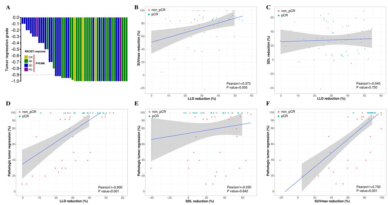
Correlation of radiological response and pathological tumor regression. (A) Pathological regression was marginally correlated with radiological response by RECIST assessment (p=0.046). (B) LLD reduction was positively correlated with SUVmax reduction of primary tumor (p=0.005). (C) LLD reduction was not correlated with SDL reduction (p=0.750). (D) LLD reduction was positively correlated with pathological tumor regression (p<0.001). (E) SDL reduction was not correlated with pathological tumor regression (p=0.842). (F) SUVmax reduction of primary tumor was positively correlated with pathological tumor regression (p<0.001). LLD, lesion longest diameter; PCR, pathologically complete response; SDL, short diameter of the largest.
A representative case of radiological response of the patient with tumor and metastatic lymph nodes was presented in figure 3. The median LLD reduction reached 31.4% (SD, 13.2%), and the median SDL reduction reached 27.9% (SD, 23.1%) revealed by post-treatment CT scan. Moreover, the median SUVmax reduction of primary tumor on post-treatment PET-CT reached 84.1% (SD, 25.3%).
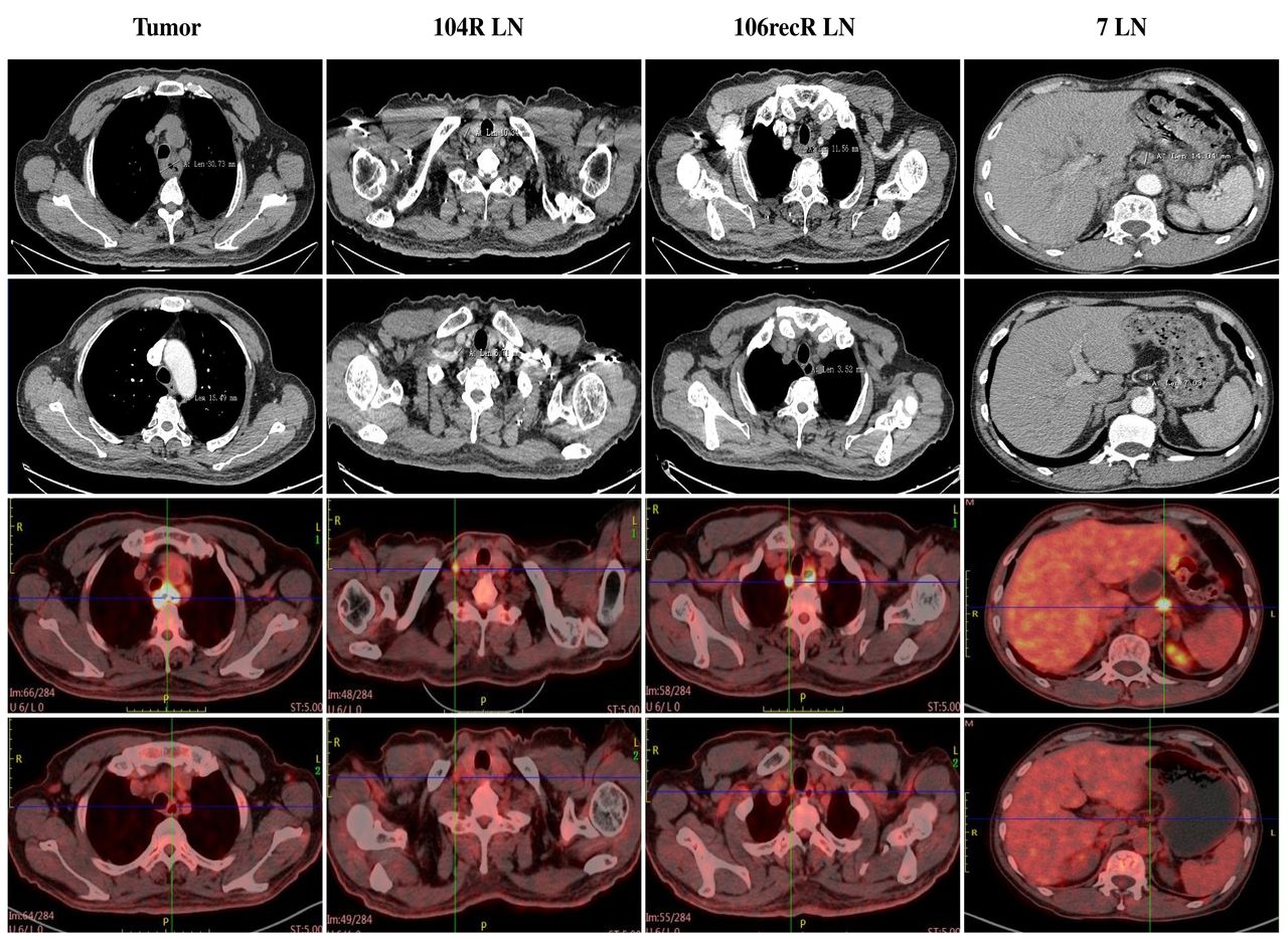
Case of radiological responses after neoadjuvant camrelizumab and chemotherapy. CT and PET-CT images before (upper row) and after (lower row) neoadjuvant treatment of patient 57 were compared. This shows the radiological images of a 67-year-old man with a stage III ESCC before neoadjuvant treatment. CT and PET-CT shows significant shrinkage and SUVmax reduction for the primary tumor and suspected lymph nodes, respectively. This patient achieved pathological regression of 95% for esophageal lesion with no residual lymph node metastasis according to surgical specimen. ESCC, esophageal squamous cell carcinoma; RLN, recurrent laryngeal nerve.
Exploratory analysis
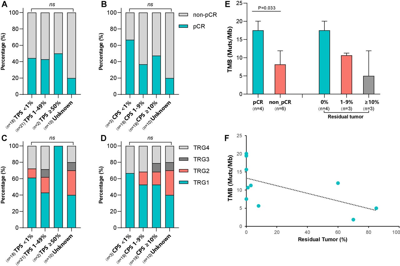
Correlation between potential biomarkers and pathological tumor regression was analyzed. There was no significant correlation between PD-L1 status and pathological response in ESCC, regardless of the assessment method of determining PD-L1 expression (figure 4A–D). Additionally, we examined the TMB of pretreatment biopsies tumor obtained from 10 patients who had adequate available tissue. All of the 10 patients who provided samples for sequencing had underwent complete tumor resection and were evaluated for tumor response. Significantly higher TMB was observed in tumors with pCR compared with tumors without pCR (p=0.033) (figure 4E). TMB levels were inversely correlated with the percentage of residual tumor (p=0.018, r=−0.752) (figure 4F). There was no significant association between the TMB levels and tumor PD-L1 expression.
Association between biomarkers and pathological regression. (A–D) PD-L1 scores and relationship to pathological regression. (E) Comparison of TMB between the PCR and non-pCR groups. (F) Distribution of TMB and the percentage of residual viable tumor cells. The dashed black line indicates the linear regression line. CPS, Combined Positive Score; PCR, pathologically complete response; TMB, tumor mutational burden; TPS, Tumor Proportion Score; TRG, tumor regression grade.
Discussion
In this multicenter, single-arm, phase II study, neoadjuvant camrelizumab in combination with chemotherapy resulted in a pCR rate of 39.2% in ESCC patients who had multi-station (≥3 stations) lymph node metastasis. Notably, this study was conducted on patients with clinical stage N2-3 disease. Given the hypothesis that a restricted radiation field has limited efficacy in high-risk patients with extensive lymph node metastasis and potential hematological metastasis, chemotherapy plus immunotherapy was used in this study. In contrast, landmark trials such as NEOCRTEC5010 trial,23 CROSS trial l,24 and FFCD 9901 trial,25 enrolled patients with clinical stage N0-1 disease. These trials administered patients with chemoradiotherapy and showed high pCR rates of 33.3%–49%, which may be attributed to improved local control with radiotherapy.
Immune checkpoint blockade is recommended for patients with advanced malignancies based on the durable anti-tumor activity and prolonged OS. The Checkmate 577 trial26 of patients with R0-resected esophageal or gastro-esophageal junction cancer with residual pathological disease also showed a significant improvement in disease-free survival in patients receiving adjuvant nivolumab compared with those receiving placebo. In the neoadjuvant setting, checkpoint blockade is deemed to eliminate micrometastasis and thus lead to superior survival by inducing system immune activation.27 Expansion of tumor-resident T cell clones in the peripheral blood was found in the neoadjuvant immunotherapy study.28 A polled analysis of neoadjuvant therapy in melanoma demonstrated pathological response were associated with improved RFS and OS.29 Here, we report short-term results, further long-term outcomes are being followed up. Similarly, a phase II study of chemotherapy followed by checkpoint blockade in patients with locally advanced ESCC reported short-term result with a pCR rate of 36.4%.30 The addition of apatinib (a VEGFR2 inhibitor) to neoadjuvant chemotherapy and camrelizumab resulted in a pCR rate of 24.1% in a phase Ib study of locally advanced ESCC.31 Although the pCR rate of 39.2% in this study is numerically higher than those in these two studies, cross-comparisons between trials need to be made with caution. Furthermore, the PALACE-1 study11 showed a higher pCR rate of 55.6% for neoadjuvant pembrolizumab plus chemoradiotherapy compared with this study. Radiotherapy and distinct patient population (35% of enrolled patients with clinical stage N0-1) could explain the difference of efficacy.
Currently, cisplatin/5-fluorouracil (PF) and carboplatin/paclitaxel (TC) are the preferred regimen of chemotherapy. PF-based regimen have been widely adopted for decades and is most commonly used regimen in Europe and North-America.32–34 The landmark CROSS trial using TC regimen with 41.4 Gy of radiation, which prompted many centers to change their standard treatment to CROSS. However, there remains no prospective head-to-head comparative study to evaluate the TC-based and PF-based regimens. Available data are retrospective studies, with small sample sizes and inconclusive results.35 36 TC-based regimen is most widely used in China because it is well tolerated by Chinese patients. As a result, TC-based regimen was chosen as the chemotherapy in this study. In addition, corticosteroids are often recommended prior to paclitaxel treatment by considering the adverse effects and dose–response relationship of paclitaxel. Several studies analyzed the association between corticosteroids and survival in patients treated with checkpoint blockade. Patients with non-small cell lung cancer receiving no less than 10 mg prednisone equivalent of corticosteroids per day prior to immunotherapy had poorer progression-free survival and OS.37 When glucocorticoids were used to manage irAEs in advanced melanoma, early high-dose glucocorticoids were associated with decreased long-term outcomes.38 However, pembrolizumab combined with carboplatin and paclitaxel or nab-paclitaxel achieved equivalent efficacy in KEYNOTE-407 trial for the first-line treatment of advanced lung squamous cell cancer.39 Nab-paclitaxel is a nanoparticle albumin-bound form of paclitaxel. It is more convenient to use than paclitaxel as it does not required pretreatment with corticosteroids. Therefore, we selected nab-paclitaxel in this study. Furthermore, this study referred chemotherapy regimen of the Keynote-407 trial, which used nab-paclitaxel (100 mg/m2, day 1, 8, 15) plus cisplatin (area under the curve of 6) every 3 weeks and resulted in an ORR of 62% with an acceptable safety profile.
Overall, the safety profile seen in this study was similar to the CROSS and NEOCRTEC5010 trials beyond irAEs, with the most common grade III AE being lymphopenia. Compared with the ESCORT-1st randomized trial,7 the RCCEP was lower in this study (26.7% vs 79.9%). The drug doses were different, two cycles of camrelizumab in this study compared with six cycles in the ESCORT-1st trial. Moreover, the median time to onset of RCCEP was 0.9 months (range 0.0–8.1) according to the product description. The ESCORT-1st trial had a longer follow-up time with a median of 10.8 months in contrast to 6.2 months in this study. Another study reported that the rate of RCCEP was only 9% after two cycles of neoadjuvant camrelizumab plus chemotherapy in locally advanced ESCC.40 Nevertheless, irAEs should be paid special attention during neoadjuvant immunotherapy. One patient developed grade 5 AE due to acute respiratory failure in this study. Thus, early warning and timely management of irAEs should be done as much as possible. No esophageal perforation bleeding was observed in our study, which had been reported in the PALACE-1 study11 and resulted in one patient death.
Immunotherapy has been reported to bring about heavy tissue reactions, which could lead to a dense and hard surgical field. However, it was not encountered in this study. The intraoperative interstitial space was largely similar to changes after neoadjuvant chemotherapy. Occasionally, tissue edema occurred, but it did not increase the operation difficulty. The mean operative time was 228 mins, which was consistent with our previous results.41 Minimally invasive technique accounted for 98% of surgical approaches and the conversion rate was extremely low with only 0.7%, which also confirmed that the regimen did not increase the operation difficulty.
In this study, we can see that pathological tumor regression was marginally associated with radiologic response by RECIST assessment. Moreover, overall tumor burden reduction, consisting of tumor size and metabolism, was closely related to pathological regression. Assessment of primary tumor response by imaging may be a potential predictor of pathological response. PD-L1 expression did not correlate to tumor response, which was in line with ESCORT-1st trial.7
Study limitations include the single-arm study design and the small number of patients. Second, although TMB level was inversely correlated with the percentage of residual tumor, the results were obtained from 10 patients whose samples were qualified for sequencing. Thirdly, the NICE regimen was demonstrated as a feasible and safe neoadjuvant treatment for locally advanced ESCC, but the long-term outcomes are still lack, especially that whether it can reduce distant metastasis. This regimen is being evaluated in an ongoing phase III randomized controlled trial (NCT05043688), comparing preoperative NICE treatment and neoadjuvant chemoradiotherapy in locally advanced ESCC.
In conclusion, NICE regimen demonstrated robust antitumor activity in locally advanced ESCC. TRAEs were generally consistent with the safety profile of neoadjuvant chemoradiotherapy. These data establish camrelizumab plus weekly chemotherapy as a promising neoadjuvant treatment for locally advanced ESCC, and further phase III randomized controlled trial is warranted.
Data availability statement
Data are available on reasonable request. Data used and analyzed during this study are available from the corresponding author on reasonable request.
Ethics statements
Patient consent for publication
Ethics approval
The study protocol was approved by the medical ethical review committee of participating institutions (No. KS1951) and conducted in accordance with the provisions of the Declaration of Helsinki and Good Clinical Practice guidelines.
Acknowledgments
The authors would like to give our heartfelt gratefulness to Prof. J. Jan B. van Lanschot (the principal investigator of CROSS trial) from the Netherlands for his suggestions to our revised manuscript. This work was supported by Jiangsu Hengrui Pharmaceutical. We are grateful to all patients and their families and all members of the collaborative group in this trial. The corresponding author had full access to all the data in the study and had final responsibility for the decision to submit for publication. All authors approved submission of the manuscript and vouch for the completeness and accuracy of the data.
References
Footnotes
Correction notice This article has been corrected to add 'Shanghai Chest Hospital' before 'Shanghai Jiao Tong University' in the affiliations of the relevant authors.
Contributors ZL conducted the study, accepted full responsibility for the study, had access to all the data and the final decision to submit for publication. JL, YY and ZL contributed equally to this study. YY, ZL, YS, HC, BY and RZ enrolled patients and collected the data. YY and ZL analyzed the data, and all authors participated in data interpretation. XF, XC, HL, LZ, YS and MZ provided the administrative support. JL, YY and ZL drafted the manuscript and all authors reviewed. The final version was approved to be submitted by all authors.
Funding ZL received funding from the National Natural Science Foundation of China (82172756) for basic research.
Disclaimer The funders had no role in study design, data collection, data analysis, data interpretation, or writing of the report.
Competing interests No, there are no competing interests.
Provenance and peer review Not commissioned; externally peer reviewed.
Supplemental material This content has been supplied by the author(s). It has not been vetted by BMJ Publishing Group Limited (BMJ) and may not have been peer-reviewed. Any opinions or recommendations discussed are solely those of the author(s) and are not endorsed by BMJ. BMJ disclaims all liability and responsibility arising from any reliance placed on the content. Where the content includes any translated material, BMJ does not warrant the accuracy and reliability of the translations (including but not limited to local regulations, clinical guidelines, terminology, drug names and drug dosages), and is not responsible for any error and/or omissions arising from translation and adaptation or otherwise.