Article Text

Abstract
Background Breast cancer(BC) is the second most common cause after lung cancer of malignant pleural effusions(MPEs),in approximately one third of all MPEs.Although,MPEs are relativity easy to be collated are still not well characterized in their cellular compositions. This opens new avenues to characterize the cellular milieu comprising the MPE, as it has the potential to be highly informative about mutational markers and immune response –ultimately guiding targeted therapy and predicting therapeutic outcomes with their study. The proposed study will characterize immune landscape of the cellular composition of MPE from patients with metastatic breast carcinoma and characterize their relationship with clinicopathologic features in these patients.
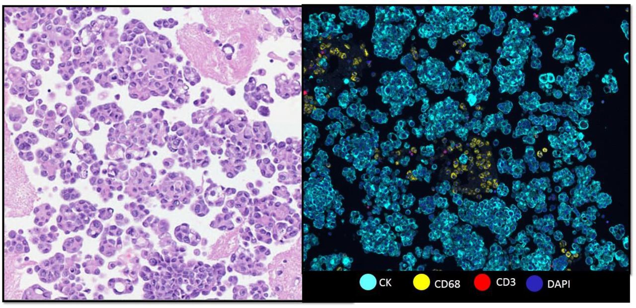
Comparison between the cell block in H-E and mIF expression CK, CD68 and CD3

Composite image in mIF expressing 8 markers. In higher magnification is possible to observe the co expression of CK+Ki67+, CK PDL1, CD3+Foxp3+ and CD3+CD8+
Results: cell phenotypes in percentage in the six cases analyzed
Clinical data of the six patients. L: left . R: right , BR : Breast cáncer, CRC: Colorrectal cáncer, NE: No evaluable , IDC : Invasive ductal carcinoma , CT: chemotherapy and BT : biotherapy
Methods Five microns thickness paraffin cell pellet blocks from six cases randomly selected of breast carcinoma MPE were stained using a quantitative multiplex immunofluorescence(mIF) panel containing 8 markers against pancytokeratin(CK), PD-L1, PD-1, CD3, CD8, Foxp3, CD68, Ki67, and DAPI (figure 1). Representative regions of interest were scanned using a multispectral scanner (Vectra Polaris) in high magnification (20x) to capture different cell populations. Markers co-expression were processed and analyzed using a quantitative image analysis software (InForm). The final results were obtained as absolute number of cells from each phenotype and were characterized with clinicopathologic features.
Results We analyzed and stained six breast cancer MPE cases with previously optimized and validated mIF panel for formalin fixed and paraffin embedded (FFPE) tumor tissues against CK, CD3, CD68, CD8, Foxp3, Ki67, PD1 and PD-L1 (figure 2). The median cellular density was 5870.53 cells. Median for each marker: CK+ was presented in 75.9% (between malignant cells and reactive mesothelial cells) in these cells the expression of Ki67 was 8% and PD-L1+ was present in 0.2%.CD3+ was 0.72% and being the cytotoxic T-cells CD3+CD8+ was 12.13% of these cells and it expression for CD3+PD1+ was in 1.14% without concomitant expression for PD-L1. The median of the macrophages CD68+ was 8.1% of the total cells (table 2).
Conclusions mIF is a promising tool to study diverse corporal effusion from different origin. Although more studies are needed, this new perspective can help us to resolve some clues and possible prognosis in advanced stages of BC.
Reference
Nicholas D T, Matthew A. S. Diagnosis and Management of Pleural Metastases and Malignant Effusion in Breast Cancer.En: Kirby I B, Edward M C, V. Suzanne K, William J. G. The Breast (Fifth Edition): Elsevier; 2018. P 934.