Article Text

Abstract
Introduction Several studies have shown clinical outcomes data that support the use of CD274 (PD-L1) copy-number (CN) gains and/or losses as a biomarker for immune checkpoint inhibitor (ICPI). Here, we present the landscape of CD274 CN changes across a large cohort of solid tumor cases and correlate these with PD-L1 protein expression by immunohistochemistry.
Methods We analyzed all cases that underwent comprehensive genomic profiling (CGP) testing at Foundation Medicine between August 2014 and June 2020. CD274 CN changes were correlated with PD-L1 expression in tumor types where there were Food and Drug Administration approved companion diagnostic (CDx) claims and the CDx assay was used to assess PD-L1 expression.
Results In all, 244 584 samples representing 290 solid tumor types were included in the study. Overall, 17.6% (42 983/244 584) had CD274 CN gains (>specimen ploidy), 44.6% (108 970/244 584) were CD274 CN neutral, and 37.9% (92 631/244 584) had CD274 CN loss. Using different CN cut offs to define CD274 positivity resulted in different prevalence estimates: ploidy +1, 17.4% (42 636/244 584); ploidy +2, 6.2% (15 183/244 584); ploidy +3, 2.2% (5375/244 584); ploidy +4, 1.1% (2712/244 584); and ploidy +8, 0.2% (434/244 584). The prevalence of CN changes and CN positivity varied based on tumor type. CD274 CN gains were significantly associated with PD-L1 positivity in NSCLC, urothelial carcinoma, breast carcinoma, cervical carcinoma, esophagus squamous cell carcinoma (SCC) and head and neck SCC (ORs 3.3, 3.0, 2.0, 4.5. 3.8, 8.4, 1.4, respectively; p<0.05) and with microsatellite instability status in only clinically relevant tumor types (gastric adenocarcinoma, colorectal adenocarcinoma, uterine endometrial adenocarcinoma, esophageal adenocarcinoma and gastroesophageal junction adenocarcinoma (OR: 5.2, 1.9, 3.2, 3.7 and 6.5, respectively; p<0.05)). Conversely, CD274 CN changes were not significantly correlated with tumor mutational burden in almost all the tumor types.
Conclusion CD274 CN changes and PD-L1 expression were highly correlated in multiple tumor types. These prevalence data on CD274 CN changes across a large cohort of different solid tumors can be used to design future clinical studies to assess whether CD274 CN changes could be a potential biomarker for ICPI.
- biomarkers
- tumor
- immunotherapy
- tumor biomarkers
Data availability statement
All data relevant to the study are included in the article or uploaded as online supplemental information. The data generated by the research that supports our article will be provided in the supplements. Due to the risk of patient reidentification, we are unable to share the raw alteration data. Academic researchers can gain access to the data in this study by filling out a study review committee form and by contacting the corresponding author. For further questions please reach out to Karen Schorr, Chief Compliance Officer, Foundation Medicine, Cambridge, Massachusetts, USA (kschorr@foundationmedicine.com).
This is an open access article distributed in accordance with the Creative Commons Attribution Non Commercial (CC BY-NC 4.0) license, which permits others to distribute, remix, adapt, build upon this work non-commercially, and license their derivative works on different terms, provided the original work is properly cited, appropriate credit is given, any changes made indicated, and the use is non-commercial. See http://creativecommons.org/licenses/by-nc/4.0/.
Statistics from Altmetric.com
Introduction
Immune checkpoint inhibitors (ICPI) have revolutionized treatment options for cancer patients, and three biomarkers are currently approved by the United States Food and Drug Administration (FDA) as companion diagnostics (CDx) for ICPI.1 These include microsatellite instability (MSI) testing, where MSI-High (MSI-H) patients with solid tumors are eligible for pembrolizumab; tumor mutational burden (TMB) testing by comprehensive genomic profiling (CGP), where solid tumor patients with TMB ≥10 mutations/megabase (mut/Mb) (TMB-High, TMB-H) are also eligible for pembrolizumab; and PD-L1 expression measured by immunohistochemistry (IHC), where PD-L1 positive tumor cells or immunocytes in certain tumor types enable the selection of ICPI such as pembrolizumab, atezolizumab or nivolumab.2–6 One promising but not as well studied biomarker for ICPI is CD274 (PD-L1) gene copy number (CN) changes.
In clinically advanced Hodgkin lymphoma, a tumor type that generally responds well to ICPI, CD274 CN gains is almost always present.7–9 In addition, in a study using CGP, CD274 amplification (defined as ploidy +4 (CN 6)) was identified in 0.7% of patients across a large cohort of diverse solid tumors, and some evidence emerged indicating that CD274 amplification is a predictor of ICPI response.10 More recently, in the SAFIR02-IMMUNO Randomized Phase II Trial, exploratory analysis has shown that CD274 amplification (defined as CN ≥5) and CN gains (CN 3–4) were predictors of durvalumab response in metastatic breast cancer.11 In non-small-cell lung cancer (NSCLC), Lamberti et al12 discovered that CD274 loss was associated with a lower response rate and progression free survival when compared with patients without CD274 loss. In another study, the authors concluded that CD274 amplification (defined as CD274 to CEP9 ratio of at least 2.0 as determined by fluorescence in situ hybridization) was associated with response to nivolumab monotherapy in NSCLC patients.13 While these studies used different methodologies to detect CD274 CN changes, overall the data showed that increases in CD274 CN were associated with better response to ICPI and decreases in CD274 CN were associated with an attenuated response to ICPI.
Currently, evidence exists in the literature supporting CD274 CN gains and losses as a promising biomarker for ICPI; however, the landscape of CD274 CN changes in different tumor types has not been well described. In addition, while a strong correlation was observed in CD274 amplification (ploidy +4 (CN 6) with CGP) and PD-L1 positivity across solid tumor types, a more nuanced study of CD274 CN and PD-L1 expression is lacking.3 12 Here, we investigated the landscape of CD274 CN changes as detected by CGP in a large cohort of diverse solid tumor cases and correlated the CD274 CN to the well-established ICPI biomarkers of PD-L1 IHC, MSI and TMB. We show that the prevalence of CD274 CN changes varied based on tumor type, and that CD274 CN changes and PD-L1 expression were highly correlated in multiple tumor types.
Materials and methods
Data collection
We analyzed all cases that underwent CGP testing at Foundation Medicine between August 2014 and June 2020. Available clinical information for the patient samples were extracted from accompanying test requisition form and pathology reports.
All specimens were assigned a diagnosis by a board-certified pathologist based on microscopic examination of a H&E-stained slide from the formalin-fixed-paraffin-embedded (FFPE) tissue, pathology report, and clinical information provided by the ordering physician.
Comprehensive genomic profiling
CGP was performed on hybridization-captured, adaptor ligation-based libraries using DNA extracted from FFPE tumor tissue in a Clinical Laboratory Improvement Amendments (CLIA)-certified and College of American Pathologists (CAP)-accredited laboratory (Foundation Medicine, Cambridge, Massachusetts, USA). The samples were sequenced for up to 324 cancer related genes and/or select gene rearrangements.14
CN alterations were detected using a comparative genomic hybridization-like method applied to next generation sequencing data.14 15 In the laboratory, each specimen was analyzed alongside a process-matched normal control (an internally validated mixture of 10 heterozygous diploid samples from the HapMap project), with custom algorithms to normalize the sequence coverage distribution across captured DNA regions. Log-ratios of normalized coverage data for exonic, intronic, and SNP targets accounting for stromal admixture, as well as genome-wide SNP frequencies, were used to generate the profiles. Using circular binary segmentation, custom algorithms further clustered groups of targets and SNP frequencies to define upper and lower bounds of genomic segments. Empirical Bayesian algorithms employed a distribution of parameters including purity and base ploidy and probability matrices were derived using different statistical sampling methodologies to fit these data. Specimen level ploidy was estimated as described by Sun et al15. Computational models were reviewed by expert analysts for each sample.14 CD274 CN gain was defined as any CN >ploidy of the specimen, CD274 CN neutral was any CN that equaled the ploidy of the specimen, and CD274 CN loss was any CN <ploidy of the specimen. We also explored prevalence rates of different CN cut offs (ploidy +1 (CN 3), ploidy +2 (CN 4), ploidy +3 (CN 5), ploidy +4 (CN 6), and ploidy +8 (CN 10)) to define CD274 CN positivity.
TMB was determined on 0.8–1.1 Mb of sequenced DNA and calculated as the number of non-driver somatic coding mut/Mb of genome sequenced.16 For TMB, we considered a TMB cut-off of at least 10 mut/Mb as TMB-H in our analysis based on the FDA pan-solid tumor CDx approval for pembrolizumab.17 MSI status was determined by analyzing 114 intronic homopolymer repeat loci for length variability, as previously described.18 MSI positivity was defined as MSI-H as per the pan-tumor approval for pembrolizumab.19
As an exploratory analysis, we hypothesized that the high rates of CD274 CN gains in cervix squamous cell carcinoma (SCC) and Head and Neck (HN) SCC could be due to human papillomavirus (HPV) infection, and therefore explored the HPV status of these patients. To determine the HPV status of the samples, we performed de novo assembly of non-human sequencing reads and BLASTn comparison against all viral nucleotide sequences in the NCBI RefSeq database were used to detect the presence of HPV genome sequences (Research Use Only).
PD-L1 IHC
PD-L1 IHC testing was run and interpreted by board-certified pathologists according to the manufacturer instructions in a CLIA-certified and CAP-accredited laboratory (Foundation Medicine, Morrisville, North Carolina) for a subset of specimens in this cohort.20 21 Specially, we examined the tumor types with a PD-L1 CDx approval: DAKO 22C3 PD-L1 assay for NSCLC (tumor proportion score cut-off ≥1), cervical carcinoma (combined positive score (CPS) cut-off ≥1), head and neck SCC (CPS cut-off ≥1), gastric/gastroesophageal adenocarcinoma (CPS cut-off ≥1), urothelial carcinoma (CPS cut-off ≥10), and esophageal SCC (CPS cut-off ≥10); and VENTANA SP142 PD-L1 assay for breast carcinoma at tumor infiltrating immune cell cut-off of 1%.22–24
Analyses
Statistical analyses were performed using R V.3.6.0 (R Foundation for Statistical Computing, Vienna, Austria) and Python V.2.7.16.25 26 Differences among categorical variables were assessed using the Fisher’s exact test. Statistical tests were two sided and multiple hypothesis testing correction was performed using the Benjamini-Hochberg procedure.
Currently, PD-L1 IHC is the gold standard for the tumor types with a PD-L1 CDx claim. We further sought to investigate the sensitivity, specificity, positive predictive value and negative predictive value of CD274 CN positivity (based on different CN cut-offs) when compared with PD-L1 IHC positivity.
Results
CD274 CN changes in various tumor types
We analyzed CD274 CN in 244 584 solid tumor samples representing 290 solid tumor types. Overall, 17.6% (42,983/244,584) had CD274 CN gains (CN >ploidy), 44.6% (108,970/244,584) were CD274 CN neutral (CN=ploidy), and 37.9% (92,631/244,584) had CD274 CN loss (CN <ploidy) (online supplemental table 1).
Supplemental material
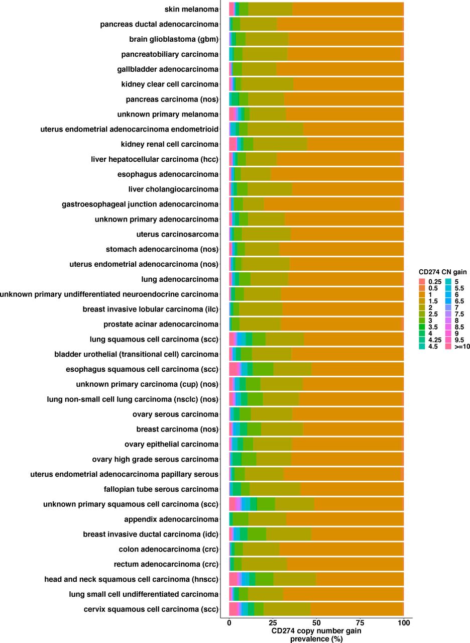
Among tumor types with ≥1000 samples, cervical SCC (31.3%), lung small-cell undifferentiated carcinoma (27.0%), and head and neck SCC (HNSCC) (25.7%) had the highest frequencies of CD274 CN gain (figure 1). Conversely, skin melanoma (8.1%), pancreatic ductal adenocarcinoma (8.5%), and glioblastoma (9.3%) had the lowest prevalence of CD274 gain. In addition, in the cohort with CD274 CN gains, we observed the highest magnitude of CN gains in tumor types with SCC morphology (figure 2). A positive correlation of CD274 CN gains with HPV infection in cervical SCC and HNSCC was observed (OR=1.4, p=0.17; OR 3.8, p=8.8×10⁻³¹). Pancreatic ductal adenocarcinoma (56.3%), gallbladder adenocarcinoma (55.5%), and cutaneous melanoma (55.4%) had the highest frequencies of CD274 CN loss. This contrasted with cervical SCC (14.4%), appendiceal adenocarcinoma (16.4%), and uterine endometrial adenocarcinoma (16.6%), which had the lowest frequencies of CD274 CN loss (figure 1).

Prevalence of CD274 copy number (CN) gains and losses in different tumor types. Cervical SCC (31.3%), lung small-cell undifferentiated carcinoma (27.0%), and head and neck SCC (25.7%) had the highest frequencies of CD274 CN gain; cutaneous melanoma (8.1%), pancreatic ductal adenocarcinoma (8.5%) and glioblastoma (9.3%) had the lowest frequencies of CD274 gain. Pancreatic ductal adenocarcinoma (56.3%), gallbladder adenocarcinoma (55.5%) and cutaneous melanoma (55.4%) had the highest frequencies of CD274 loss; cervical SCC (14.4%), appendiceal adenocarcinoma (16.4%) and uterine endometrial adenocarcinoma (16.6%) had the lowest frequencies of CD274 loss. Only tumor types with at least 1000 samples are shown.

Histogram of degree of CD274 CN gains in different tumor types. The highest levels of CN gains were observed in tumor types with SCC morphology. Only tumor types with at least 1000 samples are shown. CN, copy number.
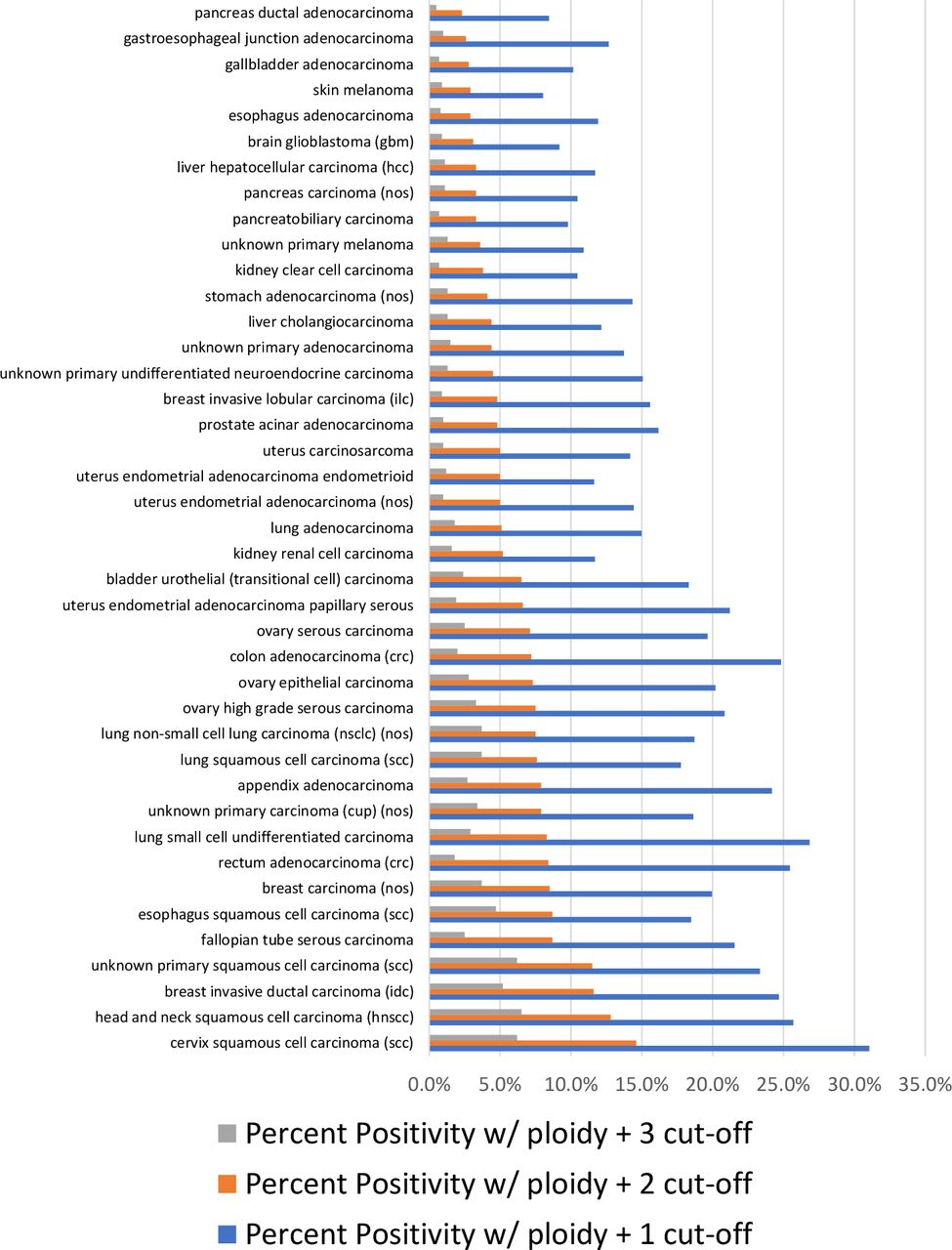
In addition, we explored the prevalence of CD274 CN positivity based on different CN cut offs. When using ploidy +1 (CN 3) as the cut-off, the overall positivity was 17.4% (42,636/244,584); when using ploidy +2 (CN 4) as the cut-off, the overall positivity was 6.2% (15,183/244,584); when using ploidy +3 (CN 5) as the cut-off, the overall positivity was 2.2% (5375/244 584); when using ploidy +4 (CN 6) as the cut-off, the overall positivity was 1.1% (2712/244 584); and when using ploidy +8 (CN 10) as the cut-off, the overall positivity was 0.2% (434/244 584). CD274 CN positivity in different tumor types also varied using these different cut-offs (figure 3 and online supplemental table 2).

Histogram showing the prevalence of CD274 copy number (CN) positivity at different CN cut-offs in different tumor types. Specially, CN cut-off at ploidy +1 (CN 3), ploidy +2 (CN 4), and ploidy +3 (CN 5) are shown here. Only tumor types with at least 1000 samples are shown.
CD274 CN correlation with PD-L1 IHC
CD274 CN gains were highly correlated with PD-L1 IHC positive status in almost all tumor types where a CDx assay was available (figure 4A). Interestingly, HNSCC had the highest OR in this comparison and gastric/esophageal adenocarcinoma which had the lowest OR (8.44, p=3.1×10⁻²; 1.41, p=8.7×10⁻²; respectively). NSCLC, urothelial carcinoma, breast carcinoma, cervical carcinoma, and esophageal SCC all had a positive and significant OR (3.29, p=3.2×10−173; 2.97, p=3.2×10⁻¹⁵; 1.96, p=1.6×10⁻¹³; 4.51, p=2.1×10⁻⁵; and 3.81, p=8.7×10⁻², respectively).

(A) OR forest plot of CD274 copy number (CN) gains and PD-L1 immunohistochemistry (IHC) positivity. CD274 CN gains were highly correlated with PD-L1 IHC positivity in almost all tumor types where a companion diagnostic assay was available. (B) Sensitivity, specificity, positive predictive value, and negative predictive value of CD274 CN positivity (defined at different cut-offs) when compared with PD-L1 IHC positivity. When compared with PD-L1 IHC, CD274 CN positivity is highly specific and has high positive predictive value. On the other hand, sensitivity and the negative predictive value is lower. Importantly, the sensitivity, specificity, positive predictive value, and negative predictive value varied depending on which cut-off we used to define CD274 CN positivity and varied depending on tumor type/PD-L1 CDx assay and scoring algorithm used. CN 3=ploidy +1, CN 4=ploidy +2, CN 5=ploidy +3, and so forth. HNSCC, head and neck SCC; NSCLC, non-small-cell lung cancer.
While CD274 CN changes were highly correlated with PD-L1 IHC status across multiple tumor types, at a population level, there was still a subset of patients in which they were not. Specifically, 4.6% (1378/29887) of the overall cohort with CD274 CN and PD-L1 IHC data had CD274 gain but were PD-L1 negative (table 1). Conversely, 23.3% (6953/29 887) had CD274 loss but were PD-L1 IHC positive (table 1).
Subset of patients in which CD274 CN gains/losses did not correlate with PD-L1 IHC positivity
When compared with PD-L1 IHC, CD274 CN positivity (at different CN cut-offs) is highly specific and has high positive predictive value (figure 4B and online supplemental table 3). On the other hand, sensitivity and the negative predictive value is lower. Importantly, the sensitivity, specificity, positive predictive value, and negative predictive value varied depending on which cut-off we used to define CD274 CN positivity and varied depending on tumor type/PD-L1 CDx assay and scoring algorithm used (figure 4B and online supplemental table 3).
CD274 CN correlation with TMB and MSI
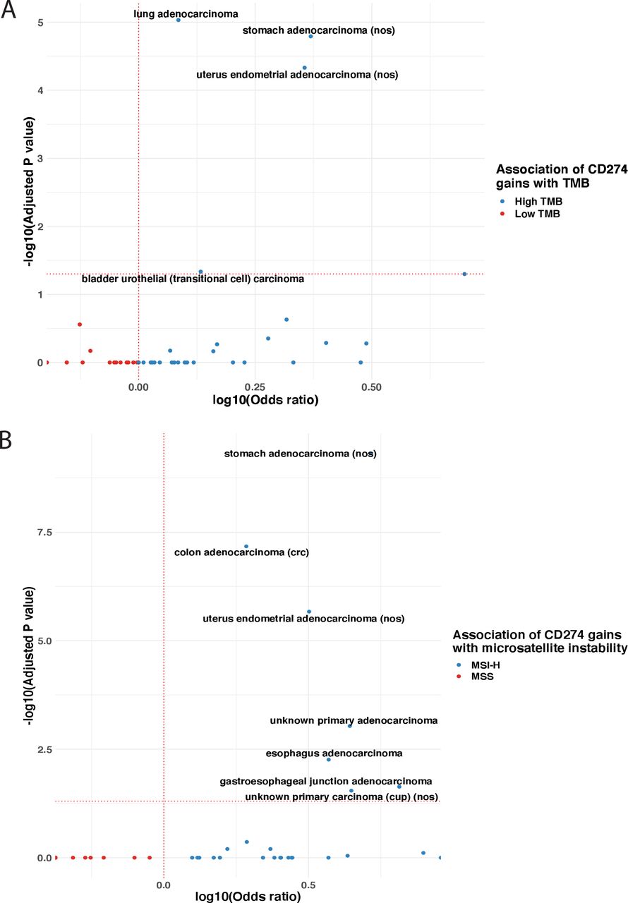
CD274 CN gains were not significantly correlated with TMB-H in almost all (98.6%, 286/290) tumor types. CD274 CN gain were significantly correlated with TMB in only four tumor types: lung adenocarcinoma, gastric adenocarcinoma, uterine endometrial adenocarcinoma, and bladder urothelial carcinoma (OR: 1.2, p=9.3×10⁻⁶; 2.3, p=1.6×10⁻⁵; 2.3, p=4.7×10⁻⁵; and 1.4, p=4.6×10⁻², respectively) (figure 5A).
(A) volcano plot depicting the association of CD274 copy number (CN) gains/losses with tumor mutational burden (TMB). The two-tailed Fisher’s exact test was used to evaluate the p values and ORs, to determine associations between CD274 CN gains/losses and TMB category (TMB-H or TMB-L) for every tumor type. The Benjamini Hochberg procedure was used to estimate the adjusted p values. Significant correlations were observed in four tumor types: lung adenocarcinoma, gastric adenocarcinoma, uterine endometrial adenocarcinoma, and bladder urothelial carcinoma. (B) Volcano plot of CD274 CN gain/losses with microsatellite instability (MSI) status. The two-tailed Fisher’s exact test was used to evaluate the p values and ORs, to determine associations between CD274 CN change and MSI category (MSI-H or microsatellite stable [MSS]), for every tumor type. The Benjamini Hochberg procedure was used to estimate the adjusted p values. In the clinically relevant MSI tumor types (gastric adenocarcinoma, colorectal adenocarcinoma, uterine endometrial adenocarcinoma, esophageal adenocarcinoma, and gastroesophageal junction adenocarcinoma), we observe a significant correlation between CD274 CN and MSI. For both A and B, only tumor types with at least 1000 samples are shown and the horizontal red dotted line represents an adjusted p value of 0.05 and the vertical dotted line represent an OR of one. MSI-H, MSI-high; TMB-H, TMB-high; TMB-L, TMB-low.
In the tumor types in which MSI is most clinically relevant (gastric adenocarcinoma, colorectal adenocarcinoma, uterine endometrial adenocarcinoma, esophageal adenocarcinoma, and gastroesophageal junction adenocarcinoma), we observed a significant correlation between CD274 CN gains and MSI-H status (OR: 5.2, p=4.9×10⁻¹⁰; 1.9, p=6.7×10⁻⁸; 3.2, p=2.1×10⁻⁶; 3.7, p=5.5×10⁻³; and 6.5, p=2.3×10⁻², respectively) (figure 5B). Most of the remaining tumor types did not have significant correlation between CD274 CN gains and MSI-H.
Discussion
In this study, we present prevalence data on CD274 CN losses, gains, and positivity (defined by different CD274 CN cut-offs) in over 240 000 patient samples across 290 solid tumor types. While Goodman et al10 previously presented data on CD274 amplification status (defined as ploidy +4) on a large cohort of patients, recent clinical data suggests that CN loss, CN gain, and amplification based on different cut offs can represent both negative and positive predictive biomarkers for ICPI response.11–13 In this study, 17.4% had CD274 CN ≥ploidy +1 (CN 3), 6.2% had CD274 CN ≥ploidy +2 (CN 4), and 2.2% had CD274 CN ≥ploidy +3 (CN 5) across tumor types, which is 25-fold, 9-fold, and 3-fold higher, respectively, than when using CD274 CN ≥ploidy +4 (CN 6), where Goodman et al10 only found 0.7% of solid tumors in their cohort as amplified. Of note, in our current cohort, we found that 1.1% (2712/244 584) had a CN ≥ploidy +4 (CN 6), which is higher than the 0.7% (843/118 187) that Goodman et al10 described. The prevalence in our study likely more accurately describes the real-world prevalence since our data set has twice the number of samples when compared with the previous study. Future clinical trials with outcomes data are needed to assess the optimal CD274 CN cut-off for a patient to be considered positive and whether CD274 CN loss is a negative predictor for ICPI response in all tumor types or only certain tumor types. Given the varied levels of CN changes in the various tumor types presented in this study, we suspect that the CN cut offs that are correlated with ICPI response will vary based on tumor type. The prevalence and diversity of CD274 CN changes in this study can serve as a basis for future clinical studies when further exploring CD274 CN changes.
Both gains and losses of CD274 were correlated with PD-L1 IHC status. This stands in contrast to genes like ROS1, where CN changes and ROS1 protein expression detected via IHC are not highly correlated.27 Instead, CD274 CN gains are more similar to ERBB2 (HER2) CN gains and HER2 protein expression in that they are correlated with each other.27 Interestingly, HNSCC CD274 CN gains had the highest correlation with PD-L1 IHC positivity. Furthermore, the highest levels of CN gains were in tumor types with SCC morphology, suggesting that CD274 CN gains could be a particularly useful biomarker for tumors with this morphology. In our exploratory analysis of HNSCC and cervical SCC, we saw a positive correlation of HPV infection with CD274 CN gain in these two tumor types. This suggests that the HPV infection likely caused the higher prevalence of CD274 CN gains in HNSCC and cervical SCC, though the exact mechanism for this remains elusive and warrants further investigation.
Lastly, when we analyzed the correlation of TMB-H with CD274 CN gains, we found that in almost all tumor types, there was no significant correlation between CD274 CN gains and TMB-H which is consistent with the findings by Yarchoan et al28 that PD-L1 expression and TMB are independent biomarkers in most tumor types. On the other hand, we saw significant correlation between CD274 CN gains and MSI-H in the tumor types where MSI is most clinically relevant, but not in most other tumor types. Importantly, subsets of patients were negative for PD-L1 IHC but had CD274 CN gain (also positive for PD-L1 IHC and had CD274 loss). Also, we saw high specificity and positive predictive value of CD274 CN positivity (with most CN cut-offs) with PD-L1 IHC positivity suggesting that CD274 CN positivity is selecting patients who are likely to respond to ICPI. On the other hand, we observed relatively low sensitivity and negative predictive value of CD274 CN positivity (at almost all CN cut-offs), meaning that CD274 CN positivity is only selecting a subset of the PD-L1 IHC positive tumors. These results in whole suggest that CD274 CN changes could be an independent positive or negative predictive biomarker for ICPI response.
Conclusion
CD274 CN changes and PD-L1 expression were highly correlated in multiple tumor types. CGP-based CD274 CN losses/gains obtained during routine clinical care could identify subsets of patients that are discordant with other known ICPI biomarkers, supporting further development of CD274 CN losses/gains as a ICPI biomarker. These prevalence data on CD274 CN changes across a large cohort of different solid tumors can be used to design future clinical studies to assess whether CD274 CN changes could be a potential biomarker for ICPI.
Data availability statement
All data relevant to the study are included in the article or uploaded as online supplemental information. The data generated by the research that supports our article will be provided in the supplements. Due to the risk of patient reidentification, we are unable to share the raw alteration data. Academic researchers can gain access to the data in this study by filling out a study review committee form and by contacting the corresponding author. For further questions please reach out to Karen Schorr, Chief Compliance Officer, Foundation Medicine, Cambridge, Massachusetts, USA (kschorr@foundationmedicine.com).
Ethics statements
Ethics approval
This study was approved by the Western Institutional Review Board Protocol No. 20 152 817.
References
Supplementary materials
Supplementary Data
This web only file has been produced by the BMJ Publishing Group from an electronic file supplied by the author(s) and has not been edited for content.
Footnotes
Contributors Conception/design: RSPH, KM, JSR. Provision of study material or patients: RSPH, KM, MM, DCP, DAM, MH, BD, GF, LAA, JSR. Collection and/or assembly of data: RSPH, KM. Data analysis and interpretation: All authors. Manuscript writing: All authors. Final approval of manuscript: all authors.
Funding The authors have not declared a specific grant for this research from any funding agency in the public, commercial or not-for-profit sectors.
Competing interests All authors of the manuscript are employees of Foundation Medicine, which is a wholly owned subsidiary of Roche and receives stock from Roche.
Provenance and peer review Not commissioned; externally peer reviewed.
Supplemental material This content has been supplied by the author(s). It has not been vetted by BMJ Publishing Group Limited (BMJ) and may not have been peer-reviewed. Any opinions or recommendations discussed are solely those of the author(s) and are not endorsed by BMJ. BMJ disclaims all liability and responsibility arising from any reliance placed on the content. Where the content includes any translated material, BMJ does not warrant the accuracy and reliability of the translations (including but not limited to local regulations, clinical guidelines, terminology, drug names and drug dosages), and is not responsible for any error and/or omissions arising from translation and adaptation or otherwise.