Article Text

Abstract
Background T cell receptor (TCR)-engineered cells can be powerful tools in the treatment of malignancies. However, tumor resistance by Human Leukocyte antigen (HLA) class I downregulation can negatively impact the success of any TCR-mediated cell therapy. Allogeneic natural killer (NK) cells have demonstrated efficacy and safety against malignancies without inducing graft-versus-host-disease, highlighting the feasibility for an ‘off the shelf’ cellular therapeutic. Furthermore, primary NK cells can target tumors using a broad array of intrinsic activation mechanisms. In this study, we combined the antitumor effector functions of NK cells with TCR engineering (NK-TCR), creating a novel therapeutic strategy to avoid TCR-associated immune resistance.
Methods BOB1, is a transcription factor highly expressed in all healthy and malignant B cell lineages, including multiple myeloma (MM). Expression of an HLA-B*07:02 restricted BOB1-specifc TCR in peripheral blood–derived NK cells was achieved following a two-step retroviral transduction protocol. NK-TCR was then compared with TCR-negative NK cells and CD8-T cells expressing the same TCR for effector function against HLA-B*07:02+ B-cell derived lymphoblastoid cell lines (B-LCL), B-cell acute lymphoblastic leukemia and MM cell lines in vitro and in vivo.
Results Firstly, TCR could be reproducibly expressed in NK cells isolated from the peripheral blood of multiple healthy donors generating pure NK-TCR cell products. Secondly, NK-TCR demonstrated antigen-specific effector functions against malignancies which were previously resistant to NK-mediated lysis and enhanced NK efficacy in vivo using a preclinical xenograft model of MM. Moreover, antigen-specific cytotoxicity and cytokine production of NK-TCR was comparable to CD8 T cells expressing the same TCR. Finally, in a model of HLA-class I loss, tumor cells with B2M KO were lysed by NK-TCR in an NK-mediated manner but were resistant to T-cell based killing.
Conclusion NK-TCR cell therapy enhances NK cell efficacy against tumors through additional TCR-mediated lysis. Furthermore, the dual efficacy of NK-TCR permits the specific targeting of tumors and the associated TCR-associated immune resistance, making NK-TCR a unique cellular therapeutic.
- cell engineering
- immunotherapy
- tumor escape
- receptors
- immunologic
- killer cells
- natural
Data availability statement
Data sharing not applicable as no datasets generated and/or analyzed for this study. Data are available upon reasonable request. All data relevant to the study are included in the article or uploaded as supplemental information. Raw data are available on reasonable request.
This is an open access article distributed in accordance with the Creative Commons Attribution Non Commercial (CC BY-NC 4.0) license, which permits others to distribute, remix, adapt, build upon this work non-commercially, and license their derivative works on different terms, provided the original work is properly cited, appropriate credit is given, any changes made indicated, and the use is non-commercial. See http://creativecommons.org/licenses/by-nc/4.0/.
Statistics from Altmetric.com
Background
The therapeutic potential of adoptive cell therapy (ACT) was actualized by the clinical success of CD19-specific chimeric antigen receptor T cell therapy (CAR-T).1 Although CD19 CAR-T is effective, disease relapse often occurs due to antigenic loss of CD192 3 emphasizing the need for alternative antigenic targets to be identified. In unmodified T cells, the T cell receptor (TCR) confers specificity and engages antigen which has been processed and presented as peptide by Human leukocyte antigen (HLA) molecules, which broadens the antigenic repertoire by including proteins from essential internal processes. The effectiveness of TCR-mediated ACT has been demonstrated by donor-lymphocyte infusion,4–6 infusions of tumor-infiltrating lymphocytes (TILs)7 or virus-specific T cells8 and TCR-engineered T cells.9–11 With the careful selection of tumor-specific targets, TCR-engineered cells have the potential to be extremely effective against malignancies.12–15 Despite this, overcoming tumor immune-evasion strategies remains a challenge for all T-cell-based immune therapies. One such strategy is HLA-class I loss, observed in patients after TIL therapy whereby unresponsive patients had dysfunctional β-2-microglobulin (B2M), required for stable HLA-class I expression.16 Furthermore, analysis of relapsed patients after immune checkpoint blockade (ICB) revealed acquired defects in antigen presentation, including B2M mutations, are contributing factors to immune resistance.17 18 Increasing TCR-mediated immune pressure on tumors, through ICB and T cell therapies, can therefore promote HLA-class I loss as a tumor immune-evasion mechanism through immunoediting.19
Recently, natural killer (NK) cells have gained interest in ACT, and thanks to advances in ex-vivo NK expansion protocols, sufficient numbers can be achieved for infusion.20 21 Once activated, NK cells share similar effector functions with T cells including production of cytotoxic granules and inflammatory cytokines. Unlike T cells, NK cell activation is independent of antigen and instead relies on a balance between activating and inhibitory signals from germline-encoded receptors.22 To prevent undesirable activation toward self-cells, NK cells express killer cell immunoglobulin-like receptors (KIRS) as well as the NKG2A/CD94 heterodimer which provide inhibitory signals on engagement with HLA-class I molecules on healthy cells. NK cells can therefore become activated by malignant cells lacking HLA-class I expression due to absence of inhibitory signals.23 The antitumor effector mechanism of KIRS has been exploited in NK-cellbased ACT through KIR mismatching. KIR-mismatched NK cells have been beneficial therapeutically and has correlated with survival advantages in patients with acute myeloid leukaemia (AML) receiving T-cell depleted allogeneic stem cell transplant (alloSCT).24 Multiple studies have since demonstrated the efficacy of allogeneic NK-ACT as a standalone therapy for the treatment of hematological and solid malignancies.25–30 Unlike T cells, allogeneic NK cells do not cause graft-versus-host-disease (GvHD), highlighting the potential for an ‘off the shelf’ cell product allowing broader patient applicability. Recently, introducing CD19 CAR into allogeneic cord blood–derived NK cells (NK-CAR) showed improved NK efficacy31 32 and results from an ongoing phase I/II clinical trial (NCT03056339) suggest NK-CAR is clinically effective and no CAR-T-associated toxicities such as cytokine release syndrome have yet been observed.33 Enhancement of NK effector function is now a focus and genetically engineering NK cells to improve efficacy, persistence and homing is part of ongoing research.31 32 34 35
In this study, we hypothesized that the combination of intrinsic, antitumor effector functions of NK cells with TCR engineering (NK-TCR) would create a therapeutic strategy to avoid TCR-associated immune resistance. Recently, TCR expression in afood and drug administration (FDA)approved NK cell line, NK-92, demonstrated TCR-mediated efficacy can be achieved in non-T- cells.36 37 Despite the advantages of a cell line as an ‘off the shelf’ cell product, NK-92 must be irradiated prior to infusion to prevent tumorigenesis, which affects the in vivo efficacy.38 Furthermore, NK-92 cells lack expression of KIRS and have low expression of NKG2A which, without further modification, would limit efficacy against malignant cells with HLA-class I loss.39 Described in this study is a two-step retroviral (RV) transduction protocol to allow functional TCR expression in primary NK cells derived from the peripheral blood of healthy donors. As a model of efficacy, we focus on the expression of a promising TCR targeting BOB1, a transcription factor highly expressed in all healthy and malignant B-cell lineages, including multiple myeloma (MM).14 We demonstrated that NK cells expressing the BOB1-specific TCR enhance NK effector functions through additional antigen-specific activation while retaining NK-mediated effector functions which could be engaged on HLA-class I loss as an immune-evasion strategy.
Materials and methods
Genetic modification of NK cells
NK-stimulator cell line K562-mbIL21-41BBL clone 41 was generated by transducing K562 with genes encoding CD137L and membrane-bound interleukin-21 (IL21) as described in a previous work40 and single cell sorted by Fluorescence-activated cell sorting (FACS). NK cells were isolated from frozen peripheral blood mononuclear cells (PBMC) using NK-cell isolation Kit (Miltenyi Biotec, Germany). In a 24-well plate, 1×10⁶ isolated NK cells were immediately co-cultured with 0.5×10⁶ irradiated (100 GY) K562-mbIL21-41BBL clone 41 in NK medium (NK-M). NK-M consisted of IMDM (Lonza, Switzerland) supplemented with 5% heat-inactivated FBS (Gibco, Thermo Fisher Scientific, USA), 5% human serum (Sanquin, The Netherlands), 1.5% 200 mM l-glutamine (Lonza), 1% 10,000 U/mL penicillin/streptomycin (Lonza), 5 ng/mL IL15 (Miltenyi Biotec, Germany) and 100 IU/mL IL-2 (Novartis, Switzerland). NK cells were then stimulated weekly with irradiated (100 GY) K562-mbIL21-41BBL at a 2:1 effector:stimulator (E:S) ratio and kept between 0.5×10⁶ and 1×10⁶ cells/mL in culture and the medium was refreshed every 2–3 days. Expansion of NK cells was calculated by live cell counts at regular timepoints after stimulation. Retroviral transduction of NK cells occurred on day 2/3 post stimulation. Transduced NK-cells were MACS (Miltenyi Biotec, Germany) enriched on day 6/7 post stimulation and immediately re-stimulated as described above. NK cells were used in effector assays between days 7 and 14 post stimulation.
Transgenic CD8 T cells
CD8 T cells (CD8T) were isolated from frozen PBMCs using CD8 microbeads (Miltenyi Biotec) and subsequently stimulated with TransAct (Miltenyi Biotec) at 10 µL/1×106 cells in NK-M without IL15. CD8T were CRISPR/Cas9 edited on day 2 with TRAC and TRBC targeting RNP as described previously.41 On day 3, CD8 T cells were retrovirally transduced with murinized tgTCR and MACS (Miltenyi Biotec) enriched on day 7 for mTCRβ expression. Enriched CD8T were cultured and used in effector assays between day 10 and day 12 post stimulation.
CRISPR/Cas9 genome editing
Ribonucleoprotein (RNP) were generated by complexing crRNA:trRNA (Integrated DNA Technologies (IDT), Coralville, Iowa, USA) with Streptococcus pyogenes (sp)Cas9 (Integrated DNA technologies (IDT)) as described previously.41 B2M-RNP were electroporated into cell lines using the NEON transfection system (Thermo Fisher Scientific, Waltham, Massachusetts, USA) using transfection settings: UM9 (1600 V 10 ms 3x pulses), BV-ALL (1550 V 10 ms 3x pulses) and EBV-LCL (1500 V 20 ms 2x pulses). B2M-KO cells were FACS sorted according to loss of HLA-class I.
B2M crRNA sequence42: 5′’-GGCCACGGAGCGAGACAUCUGUUUUAGAGCUAUGCU-3′.
In vitro effector assays
Cytotoxicity
A total of 1250 target cells were labeled with 100 µCi Na251CrO4 for 1 hour at 37°C, and co-cultured with NK or T cells at multiple effector-to-target (E:T) ratios in 100 µL IMDM supplemented with 10% Fetal bovine serum (FBS). After 6 hours of incubation at 37°C, 25 µL of supernatants was harvested). 51Cr release was measured on a 2450 Microbeta2 plate counter (PerkinElmer, Waltham, Massachusetts, USA). % cytotoxicity was calculated as follows: (Test sample 51Cr release minus the spontaneous 51Cr release)/ (maximum 51Cr release minus the spontaneous 51Cr release)×100. TCR-dependent killing was calculated as the difference in killing between NK/CD8T:BOB1 and NK:TCRneg/CD8T:MOCK at the highest E:T ratio.
Degranulation and cytokine production
Effector cells were stimulated with phorbol myristate acetate (PMA) (5 ng/mL) and ionomycin (400 ng/mL) or co-cultured with stimulator cells at an E:S ratio of 1:4 in 100 µL NK-M(IL15) in the presence of anti-CD107a. After 1 hour of incubation at 37°C, Brefeldin-A (5 µg/mL) was added and subsequently incubated for 12–14 hours. Cells were then stained with zombie-aqua (Biolegend) followed by cell surface antibodies (anti-CD56(NK)/anti-CD8(CD8)) and subsequently fixed for 12 min in 1% paraformaldehyde and permeabilized with 0.1% saponin for 20 min at 4°C. Intracellular antibody staining was then performed in 0.1% saponin for 20 min at 4°C and analyzed by FACS.
In vivo anti-myeloma efficacy
On day 0, non-irradiated female NOD.Cg-Prkdc(scid)Il2rg(tm1Wjl)/SzJ (NOD scid gamma, NSG) mice (The Jackson Laboratory, Bar Harbor, Maine, USA) were infused with luciferase positive UM9 MM cells alone or simultaneously with NK cells in 200 µL of PBS supplemented with 0.5 µg human GMP-grade IL15 (Miltenyi Biotec Bergisch Gladbach, Germany). Tumor cells and NK cells were prepared separately and mixed directly before infusion. About 0.5 µg human GMP-grade IL15 (Miltenyi Biotec) was then infused via intraperitoneal injection (200 µl) every 2–3 days for 3 weeks. To monitor tumor growth, mice were anesthetized with 3%–4% isoflurane after they received an intraperitoneal injection of 200 µL 7.5 mM D-luciferine (Cayman Chemical Company, Ann Arbor, Michigan, USA). Whole body bioluminescence images were obtained using a CCD camera (IVIS spectrum, PerkinElmer, Waltham, Massachusetts, USA). We used Animal Research: Reporting of In Vivo Experiments (ARRIVE) reporting guidelines.43
Flow cytometry
FACS analysis was performed on either the LSRII (BD Biosciences, New Jersey, USA) or Fortessa X20 (BD Biosciences), and data were analyzed using flowjo V.10 software (TreeStar, Oregan, USA). For each stain, 0.05×10⁶ NK cells were aliquoted into 96-well v-bottom plates, washed and incubated with 10 µL antibody mixes for 20 min at 4°C. For tetramer binding experiments, NK cells were first incubated with 10 µL PE-labeled pMHC tetramers for 20 min at 4°C, followed by antibody staining.
Statistical analysis
Statistical analysis was performed using GraphPad Prism7 software. When comparing two groups, statistical significance was calculated using unpaired t-test. In vivo efficacy was calculated using one-way analysis of variance with Tukey’s multiple comparison test on log-transformed data. Kaplan-Meier curve was used to depict survival with Gehan-Breslow-Wilcoxon test.
Results
Generation of TCR expressing NK cells following a two-step retroviral production protocol
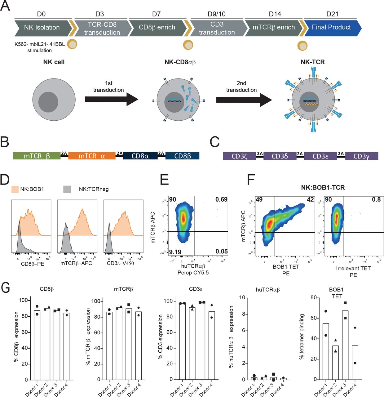
Here, we genetically engineered NK cells using RV to generate a final cell product containing TCR expressing NK cells (NK-TCR) (figure 1A). NK cells were sourced from the peripheral blood of healthy donors and stimulated weekly using a modified NK-sensitive K562 cell line, expressing a membrane-bound form of IL21 and co-stimulatory receptor 41BBL (K562-mbIL21-41BBL) (figure 1A).20 Functional TCR expression is challenging in NK cells because all missing components must be introduced alongside the TCRαβ chains, including CD8αβ co-receptor and the CD3ζγεδ signaling chains. To reduce the number of transductions, we first introduced TCRαβ and CD8αβ in one RV construct and transduction efficiency was measured by cell surface expression of CD8β, as without CD3, the TCR cannot reach the cell surface (figure 1B and online supplemental figure S1A). Introduction of BOB1-specifc TCR-CD8 resulted in efficient and reproducible CD8β expression frequencies (BOB1-TCR 34%±15.7%, mean±SD) (online supplemental figure S1A) which could be enriched for CD8β expression. Enriched CD8βpos NK cells were subsequently transduced with the four invariant chains of the CD3 signaling complex in a separate RV construct to permit cell surface expression of the introduced TCRαβ (figure 1C and online supplemental figure S1B). In these experiments, each transgenic TCR (tgTCR) was murinized (mTCR) to allow distinction between any contaminating T cells present in the culture which may influence functional data. Transduction efficiencies were consistent resulting in the expression of mTCRβ (BOB1-TCR 34%±13%) (online supplemental figure S1B), which were enriched before a final stimulation and further expansion. To test reproducibility, NK-BOB1 tgTCR cell products (NK:BOB1) were generated from four donors and repeated twice for each donor. High expression of CD8β, mTCRβ and CD3ε was repeatedly observed in NK-TCR cell products and NK-TCR specificity was confirmed using peptide-MHC tetramers (figure 1D–G). All NK:BOB1 remained negative for human TCRαβ, indicating any residual T cells present at isolation did not expand in vitro (figure 1E and G). Following this stepwise method, pure NK-TCR was generated from multiple healthy donors, within 21 days and total fold expansion of 4385±2026 SEM was observed (online supplemental figure S2). Furthermore, this protocol could be easily adapted to express different clinically relevant TCRs such as CMV-specific or PRAME-specific TCRs (online supplemental figure S3 and S4A–E).
Supplemental material

Generation of TCR expressing NK cells following a two-step retroviral production protocol. (A) Schematic of the 21-day production protocol to generate TCR expressing NK cells (NK-TCR). Retroviral construct design for (B) TCR linked via 2A sites to CD8ab (TCR-CD8) and (C) CD3ζδεγ invariant chains linked via 2A sites. (D) Representative histograms of CD8β, mTCRβ, CD3ε and human TCRαβ expression in a final NK-TCR cell product expressing BOB1-specific TCR (NK:BOB1) or NK cells without TCR expression (NK:TCRneg) on day 21. Representative FACS plot of NK:BOB1 stained for (E) mTCRβ and human (hu) TCRαβ expression and with (F) BOB1 or an irrelevant pMHC tetramer on day 21 post isolation. (G) Summary of expression frequencies of CD8β, mTCRβ, CD3ε, huTCRαβ expression and BOB1-specific tetramer binding frequencies on day 21 of NK:BOB1 cell products generated from four different donors. NK:BOB1 was generated twice for each donor. Means and individual values are depicted. NK, natural killer; TCR, T cell receptor.

NK-TCR elicit antigen-specific cytotoxicity against tumor targets. NK cells expressing the HLA-B*07:02 restricted, BOB1-specific TCR (NK:BOB1) or negative for TCR expression (NK:TCRneg) were co-cultured, at multiple E:T ratios, with 51Cr labeled (A) K562-mbIL21-41BBL or HLA-B*07:02±EBV LCLs, (B) HLA-B*07:02+fibroblasts negative for BOB1 expression, (C) HLA-B*07:02+B ALL cell line (BV-ALL) or HLA-B*07:02+ multiple myeloma cell line UM9 and leukapheresis samples from HLA-B*07:02+ patients with (D) B-cell acute lymphocytic leukemia (B-ALL) or (E) B-cell chronic lymphocytic leukemia (B-CLL). Each patient material had >70% malignant cells present and was untreated. (A)–(E): Error bars represent mean and SD of technical triplicates. (F) Summary of % cytotoxicity at the highest E:T ratio (5:1) from four NK cell products generated from different donors. Statistical test used was paired t-test. E:T, effector:target ratio; NK, natural killer; TCR, T cell receptor.

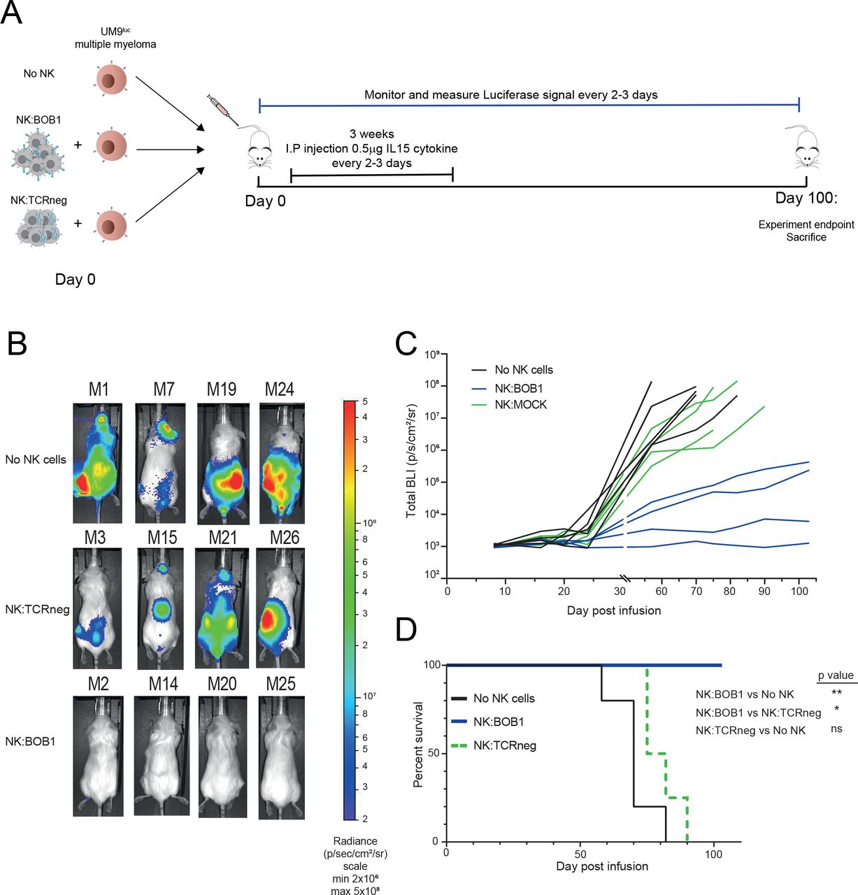
NK-TCR increases survival of treated mice in a preclinical model of multiple myeloma. (A) Schematic of experiment setup, NOD scid gamma (NSG) mice were infused intravenously with 0.4×10⁶ HLA-B*07:02+, luciferase-expressing UM9 multiple myeloma cell line alone (No NK, n=5) or simultaneously with 6×10⁶ NK cells expressing BOB1-TCR (NK:BOB1, n=4) or TCR negative NK cells (NK:TCRneg, n=4) in the presence of human IL15 cytokine. Mice were injected every 2–3 days with 0.5 µg IL15 for 3 weeks. For measurement, mice were injected intraperitoneally with luciferin and measured for bioluminescence. (B) Bioluminescent images of mice at day 70. (C) Tumor burden as measured by bioluminescence of UM9 over time. Each line represents an individual mouse . (D) Kaplan-Meier survival curve of mice receiving No NK cells, NK:BOB1 and NK:TCRneg. Remaining mice were sacrificed on day 100 at experiment end. Differences between survival was analyzed using Gehan-Breslow-Wilcoxon test. IL, interleukin; NK, natural killer; TCR, T cell receptor.

Dual-targeting characteristics of NK-TCR enhanced overall cytotoxicity against tumor targets. Alongside NK:BOB1, CD8 T-cells (CD8T), with CRISPR-mediated knock out of endogenous TCRαβ chains, were transduced to express murinized BOB1-specific TCR (CD8T:BOB1) or mock transduced with murinized CMV-specific TCR (CD8T:MOCK). (A) Representative FACS plot and summary data of mTCRβ and huTCRαβ expression in NK:BOB1 (D7 post stimulation) and CD8T:BOB1 (D10 post stimulation) generated from the same three healthy donors. (B) Overall expression frequencies and geometric mean of mTCRβ expression in CD8T:BOB1 and NK:BOB1. Error bars represent mean and SD from biological replicates. (C) % cytotoxicity of NK:BOB1 and CD8T:BOB1 against HLA-B*07:02 CD8T target cells, exogenously loaded with BOB1(B7)-peptide (APAPTAVVL) at a 10:1 E:T ratio. Each symbol represents a different donor and error bars represent mean and SD of biological replicates. Statistical test used was unpaired t-test. (D) TCR-dependent killing of NK:BOB1 and CD8T:BOB1 calculated as the difference between NK:BOB1\NK:TCRneg and CD8T:BOB1\CD8T:MOCK cytotoxicity at the highest E:T ratio. POU2AF1 encodes the BOB1 protein. (E) Combined data from multiple donors depicting the % cytotoxicity of NK:BOB1 (n=4), NK:TCRneg (n=4), CD8T:BOB1 (n=3) and CD8T:MOCK (n=3) co-cultured with HLA-class I negative K562, HLA-B*07:02±EBV LCLs, HLA-B*07:02+B ALL cell line (BV-ALL) and HLA-B*07:02+multiple myeloma cell line UM9. (A, B, D, E) Mean and SD of biological replicates are depicted, comparisons were made using unpaired t-tests. (C) Mean and SD of technical triplicates are depicted for four donors. E:T, effector:target ratio; NK, natural killer; TCR, T cell receptor.
NK-TCR express a diverse array of receptors required for NK cell activation
The phenotype of final NK-TCR cell products revealed high frequency expression of activation receptors CD2, DNAM1, CD16, NKG2D (online supplemental figure S5). Natural cytotoxicity receptors (NCRs) NKp30 and NKp46 were also highly expressed, whereas NKp44 was expressed on only 29%±3.8% (mean±SD) of NK:BOB1 (online supplemental figure S5). Additionally, high frequency expression of inhibitory receptor NKG2A was repeatedly observed in NK:BOB1 (93.5%±3.3% mean±SD) and the activating receptor NKG2C was found on a small fraction of cells (12.8%±4.8% mean±SD). KIR expression was similarly expressed on a small percentage of cells indicating the expansion protocol did not select for a particular KIR expressing population (online supplemental figure S5). As a negative control, TCR negative NK cells (NK:TCRneg) were expanded in parallel to NK:BOB1 from the same healthy donors. The phenotype of NK:TCRneg did not significantly differ except for an increased frequency of NKp44 expressing cells (54.3%±2.2% mean±SD), suggesting they were more activated, and lower CD28 expression. This expression profile suggested NK-TCR can be activated via many receptor pathways which permit antitumor effector functions.
NK-TCR elicit potent HLA-dependent, antigen-specific cytotoxicity against tumor targets
Functional capabilities of our generated NK-TCR cell products were investigated on days 21–24 without further stimulation. We validated the NK-mediated cytotoxic potential of NK:BOB1 and NK:TCRneg against HLA-class I negative K562 cell line which was always equally potent and therefore no difference was observed when BOB1-TCR was expressed (figure 2A). To explore antigen-specific killing, we designed a panel of EBV-LCLs, two endogenously expressing the BOB1 T-cell epitope in HLA-B*07:02 and two negative for HLA-B*07:02. NK:TCRneg demonstrated low levels of killing against all four EBV-LCLs representing the background NK-mediated cytotoxicity (figure 2A). In contrast, NK:BOB1 demonstrated increased killing of HLA-B*07:02+ but not HLA-B*07:02-EBV-LCLs indicative of TCR-mediated lysis (figure 2A). We confirmed antigen dependence using HLA-B*07:02+ fibroblasts which were negative for BOB1 antigen and did not induce TCR-mediated cytotoxicity (figure 2B). The expression of TCR on NK:BOB1 also increased cytotoxicity against HLA-B*07:02+ cell lines representing B-cell acute lymphoblastic leukemia (B-ALL) and MM which were previously insensitive to NK-mediated cytotoxicity (figure 2C). To expand on this further, NK:BOB1 was investigated for its ability to lyse HLA-B*07:02+ leukapheresis samples from patients with B-cell chronic lymphocytic leukemia (B-CLL) or B-ALL. All samples contained >70% malignant cells and variable levels of NK-mediated cytotoxicity were demonstrated by NK:TCRneg against each of the malignancies (figure 2D and E). As before, increased cytotoxicity was demonstrated by NK:BOB1 and the benefit of NK-TCR was more pronounced when the primary malignancy did not induce an NK-mediated response (figure 2D and E). This TCR-mediated cytotoxicity was consistently demonstrated by NK-BOB1 cell products from multiple donors (figure 2F). Furthermore, antigen-specific killing was not restricted to the BOB1-TCR, and NK-TCR expressing PRAME-specific TCR (NK:PRAME) or CMV-specific TCR (NK:CMV) were also able to specifically lyse antigen-expressing target cells (online supplemental figures S4F and S6).
NK-TCR enabled improved efficacy in a preclinical in vivo model of MM
Next, we investigated if TCR-mediated activation of NK:BOB1 would improve efficacy of NK-cell therapeutics in vivo. NSG mice were injected with HLA-B*07:02+MM cell line UM9, previously shown to have low NK-mediated activity (figure 2). NK:BOB1 and NK:TCRneg were generated from a KIR-matched healthy donor and co-injected with UM9 in the presence of IL15. Efficacy was measured by tumor outgrowth and NK:BOB1 demonstrated delayed tumor outgrowth and increased overall survival compared with NK:TCRneg and untreated mice (figure 3). These data validated the in vitro results and demonstrated that TCR expression in NK-TCR permits an additional activation pathway that ultimately improves NK-cell therapy efficacy in vivo.
The antigen-specific cytotoxicity of NK-TCR is comparable to CD8 T cells expressing the same TCR
To further understand the potency of NK-TCR, we compared cytotoxicity relative to CD8 T cells (CD8T) expressing the same TCR. To remove the influence of the endogenous TCR on tgTCR expression, the endogenous TCRαβ chains of CD8T were deleted using CRISPR/Cas9, as described previously.41 As expected, NK:BOB1 demonstrated higher frequency of single positive mTCRβ+ cells (figure 4A). However, despite the presence of a residual human TCRαβ+ cell population co-expressing mTCRβ in CD8T:BOB1 (figure 4A), the overall frequency and expression of mTCRβ+ cells was comparable between NK:BOB1 and CD8T:BOB1 (figure 4B).
Next, we conducted a peptide titration of the BOB1 epitope using BOB1-negative HLA-B*07:02+CD8T as target cells which revealed CD8T:BOB1 were more cytolytic toward targets cells presenting low concentrations of exogenously loaded antigen, compared with NK:BOB1 (figure 4C). This was also apparent for CD8T:CMV when tested against peptide-loaded HLA-A*02:01+EBV LCLs; however, here NK:CMV demonstrated enhanced cytolytic activity against targets exogenously loaded with higher concentrations of peptide (online supplemental figure S7). It is known that BOB1-peptide is less readily exogenously loaded onto target cells,14 so we proceeded to investigate antigen-specific cytotoxicity of NK:BOB1 and CD8T:BOB1 against our panel of HLA-B*07:02+B cell targets endogenously expressing BOB1. Here, TCR-dependent killing by NK:BOB1 and CD8T:BOB1 was calculated to be similar (figure 4D), reflecting the comparable levels of mTCRβ+ expression (figure 4B). However, higher overall cytotoxicity, against HLA-B*07:02+B cell targets endogenously expressing BOB1, was demonstrated by NK:BOB1 (figure 4E). The combination of NK-mediated and TCR-mediated effector actions of NK-TCR therefore offers an advantage over other TCR-engineered cell therapeutics by enhancing cytolytic efficacy via additional NK-mediated tumor targeting signals.
Multiple antigen-specific effector functions are elicited by NK-TCR
To understand the functional capabilities of NK:BOB1 further, we analyzed degranulation and cytokine production of NK:BOB1 and CD8T:BOB1 cell products. All cell products were capable of degranulation, as measured by CD107a, and production of tumor necrosis factor-α (TNFα) and interferon-γ (IFN-γ) inflammatory cytokines, except for one CD8T donor that produced low levels of cytokine (online supplemental figure S8). NK-mediated responses were demonstrated by NK:BOB1 and NK:TCRneg which, unlike CD8T, degranulated and produced TNFα and IFN-γ in response to K562 (figure 5). Antigen-specific degranulation and cytokine production were also demonstrated by NK:BOB1 and CD8T:BOB1 after stimulation with HLA-B*07:02+B cell targets (figure 5). These findings were not limited to the BOB1-TCR and NK:CMV and CD8T:CMV also showed antigen-specific responses (online supplemental figure S9). Interestingly, CD8T:BOB1 demonstrated significantly increased degranulation against MM cell line UM9 (figure 5A). This trend was also observed for cytokine production, and suggests the effector functions of NK-TCR may be influenced by the target cell itself (figure 5B,C). Nonetheless, these data demonstrate that NK-TCR can evoke multiple, antigen-specific effector functions.
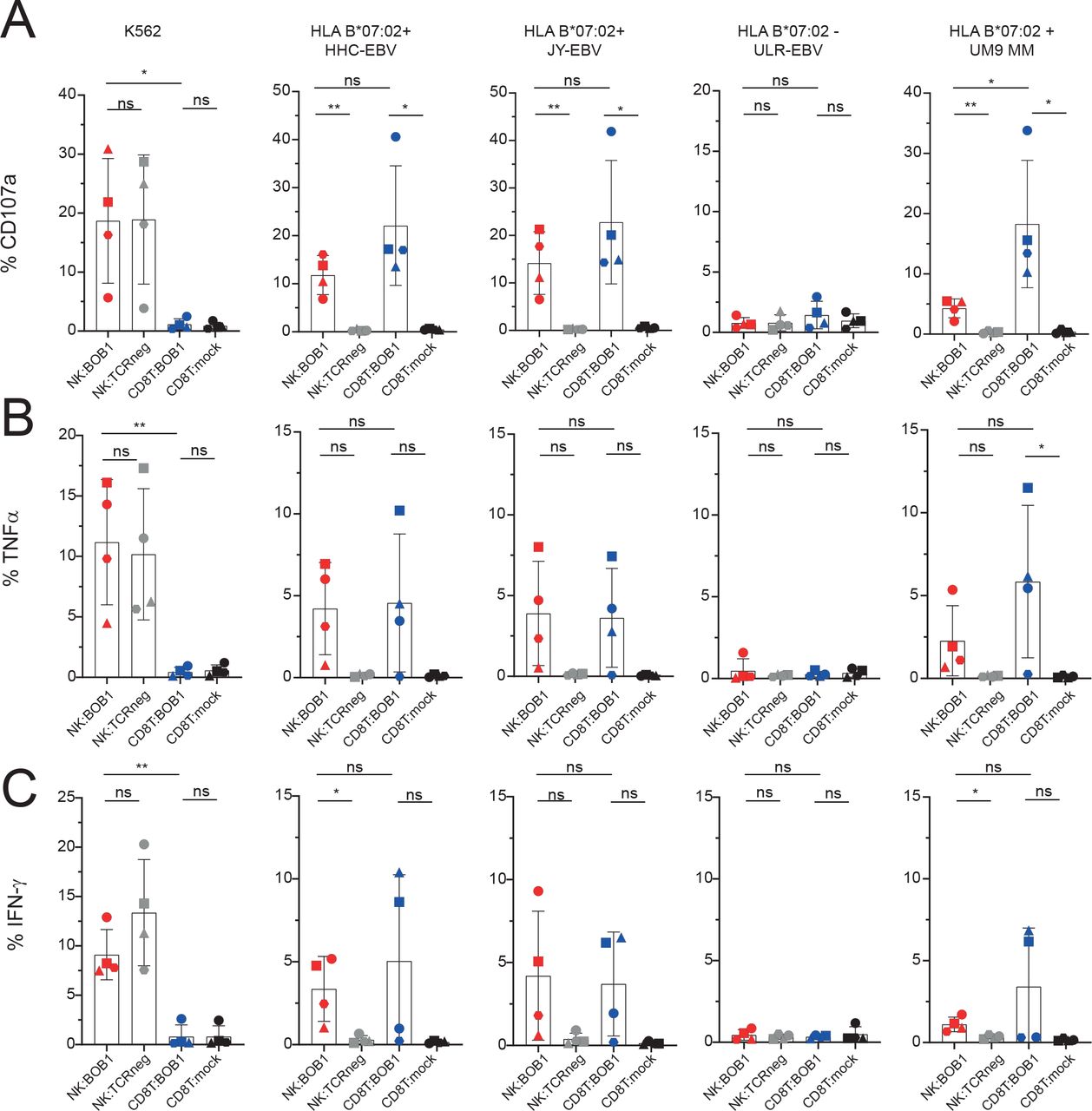
NK-TCR demonstrate antigen-specific degranulation and cytokine production. CD8T expressing BOB1-TCR or CMV-TCR (day 10 (D10) post stimulation) and NK cells expressing BOB1-TCR or TCR negative NK cells (day 7 post stimulation) were co-cultured overnight with HLA-class I negative K562, HLA-B*07:02±EBV LCLs, HLA-B*07:02+B ALL cell line (BV-ALL) and HLA-B*07:02+multiple myeloma cell line UM9 in the presence of Brefeldin-A and anti-CD107a. After 12–14 hours of incubation, cells were stained for inflammatory cytokines TNFα and IFN-γ and assessed by FACS. Depicted is the combined data of cell products derived from different donors for (A) CD107a, (B) TNFα and (C) IFN-γ expression. Live NK cells were gated on CD56 expression and live CD8T were gated on CD8 expression. Positive cells were gated according to unstimulated. Target cells were td-tomato positive and were removed from analysis. Each symbol represents a different donor and error bars represent mean and SD of biological replicates. Statistical test used was unpaired t-test. IFN, interferon; natural killer; TCR, T cell receptor; TNF, tumor necrosis factor.
NK-mediated effector functions of NK-TCR can be engaged upon HLA-class I loss on tumor targets
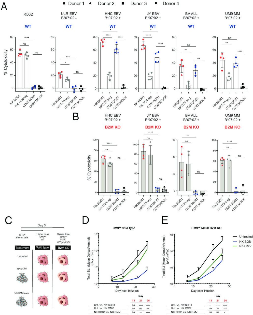
Finally, we modelled HLA-class I loss in tumors by generating HLA-B*07:02+EBV LCLs, B-ALL and MM cell lines with β-2-microglobulin knock-out (B2M KO). B2M KO prevented cell surface expression of HLA-class I molecules in all cell lines (online supplemental figure S10). B2M KO or wild-type cells were then co-cultured with NK:BOB1, NK:TCRneg, CD8T:BOB1 and CD8T:MOCK. In this model of HLA-class I loss, CD8T:BOB1 killed the wild-type cells but was unable to kill the B2M KO cells due to absence of TCR-specific epitope at the cell surface (figure 6A,B). In contrast, NK:BOB1 and NK:TCRneg demonstrated equally potent killing of B2M KO cells due to activation of NK-mediated cytotoxicity (figure 6A,B). As demonstrated previously, only NK:BOB1 was able to kill wild-type cells representing TCR-mediated killing.
NK-TCR demonstrate NK-mediated killing of tumor targets with HLA-class I loss. NK cells expressing the BOB1-specific TCR (NK:BOB1) or TCR negative NK cells (NK:TCRneg) and CD8T expressing BOB1-TCR (CD8T:BOB1) or CMV-TCR (CD8T:MOCK) were co-cultured with HLA-B*07:02+tumor targets which were either (A) wild type or (B) B2M KO. Depicted is the combined cytotoxicity data at 10:1 E:T ratio by cell products derived from four different healthy donors. Each symbol represents a different donor and error bars represent mean and SD of biological replicates. Statistical test used was unpaired t-test. (C) Schematic representation of high-dose tumor burden experiment. (D) NOD scid gamma (NSG) mice were infused intravenously with a total 0.8×10⁶ (higher dose) UM9luc alone (untreated, n=4) or simultaneously with 6×10⁶ NK:BOB1, n=5 or CMV-TCR expressing NK cells (NK:CMV n=4). (E) NSG mice were infused intravenously with a 50/50 mix of 0.4×10⁶ wild-type UM9luc and 0.4×10⁶ UM9luc B2M KO cells alone (untreated, n=4) or simultaneously with 6×10⁶ NK:BOB1, n=7 or NK:CMV n=6. IL15 was infused every 2–3 days for 3 weeks. Experiment endpoint was 6 days after IL15 withdrawal. Error bars represent mean and SD and statistical analysis was calculated using one-way analysis of variance with Tukey’s multiple comparison. B2M KO, β-2-microglobulin knock-out; E:T, effector:target ratio; IL, interleukin; NK, natural killer; TCR, T cell receptor; WT, wild type.
Next, we investigated NK-TCR efficacy in vivo against a decreased NK:tumor cell ratio by doubling the number of UM9 infused alongside NK-TCR. As before, KIR-matched NK-cells were isolated from a healthy donor and as a negative control, the CMV-TCR was also expressed in NK cells in parallel to the BOB1-TCR (figure 6C). Again, NK:BOB1-treated mice demonstrated reduced tumor outgrowth of wild-type UM9 compared with untreated mice, while NK:CMV-treated mice did not indicating presence of a non-specific TCR does not enhance NK activity (figure 6D). In parallel to this, mice were similarly infused with a 50/50 mix of UM9 wild-type and UM9 B2M KO cells alongside NK-TCR to determine efficacy against tumors with heterogenous HLA-class I expression (figure 6E). In contrast to UM9 wild-type mice, NK:CMV significantly reduced tumor outgrowth compared with untreated mice, demonstrative of NK-mediated targeting of HLA-class I negative tumor cells (figure 6E). Furthermore, NK:BOB1 demonstrated an enhanced effect as both HLA-class I negative and positive cells can be targeted (figure 6E). These data demonstrate the NK-mediated effector functions of NK-TCR can also be engaged in the absence of TCR-specific epitope following loss of HLA-class I.
Discussion
The data presented in this study demonstrate the enhancement of NK effector function by genetically engineering primary NK cells to express a TCR. We focused on the expression of a TCR specific for a B-cell-restricted antigen, BOB1, and high expression of BOB1-TCR was repeatedly observed in modified NK cells. Furthermore, our protocol can be used to express different TCRs targeting many tumor types (online supplemental figures S3 and S4). NK-TCR permits an alternative NK-activation mechanism against malignancies which are insensitive to NK-mediated attack. To date, autologous NK-ACT has not shown clinical efficacy and inhibition by self-HLA is likely to contribute to its ineffectiveness.44 45 Equipping autologous NK cells with tumor-specific TCR may therefore override inhibition signals and boost therapeutic efficacy, as similarly observed by NK cells expressing CAR. We demonstrated targets cells with high HLA-class I expression (online supplemental figure S10) elicited some NK-mediated cytotoxicity, suggesting alternative NK-activation pathways were engaged to override inhibition, and additional TCR-mediated activation enhanced this effect further (figures 4E and 6A). Furthermore, the cumulative effect of NK-mediated and TCR-mediated activation increased overall cytotoxicity against tumor targets endogenously expressing antigen when compared with tgTCR expression in T cells (figure 4E). The increased potency of NK-TCR permitted by dual-targeting actions could be beneficial in clinical contexts in which NK cells are already associated with antitumor effects, such as alloSCT. Graft verse leukemia (GvL) effects are described in patients transplanted with alloreactive KIR mismatched NK cells.24–26 46 Our data infers that tgTCR expression in infused NK cells could enhance this effect. Another application could be early infusion of allogeneic NK cells expressing virus-specific and tumor-reactive TCR following T-cell-depleted alloSCT. This would potentially allow preservation of GvL effects and protection against harmful viral re-activities in the absence of T cells, without the risk of GvHD.8
In this study, antigen-specific cytotoxicity against endogenous antigen was comparable between CD8T:BOB1 and NK:BOB1; however, exogenous loading of BOB1 peptide onto targets resulted in higher levels of cytotoxicity by CD8T:BOB1 (figure 4C). One explanation could be due to the inefficient loading of exogenous BOB1 peptide onto HLA-B*07:02 at the cell surface, as demonstrated previously.14 This results in lower levels of BOB1 peptide on exogenously loaded cells compared with B-cell targets that endogenously process and present the BOB1 epitope. NK activation is complex and, unlike CD8T, is influenced by a combination of inhibitory and activation signals. The expression levels of these signals combined with low expression of BOB1 antigen may prevent NK-cell activation thresholds being met. Understanding the effect of NK-mediated activation thresholds on NK-TCR function would be an interesting study. It is also important to note that direct functional comparisons between NK-TCR and CD8T, either in vivo or in vitro, should be made with caution as they are distinct cell types with different capabilities for activation, serial-targeting and persistence.
To our knowledge, this is the first study demonstrating TCR expression in primary NK cells derived from the peripheral blood (PB) of healthy donors. To date, functional TCR expression in non-T cells has been restricted to the NK-cell line, NK-92,36 37 largely owing to low transduction efficiencies observed in primary NK cells. As demonstrated in this study, improvements to NK expansion protocols have now enabled efficient retroviral transduction of primary NK cells (online supplemental figure S1). PB offers a readily available source of mature NK cells with a potent cytotoxic profile, leading to their use in multiple clinical studies.47 Here, PB-derived NK-TCR demonstrates multiple antigen-specific effector functions including cytotoxicity, degranulation and inflammatory cytokine production (figure 5). In allogeneic settings, alternative NK sources such as cord blood (CB) or hematopoietic stem cells have also been used due to the reduced risk of T cells being present in the graft.34 48 In this study, T cells present after NK isolation did not expand on stimulation and were absent from final NK-TCR cell products (figure 1). Although not extensively studied, we have demonstrated that functional TCR could also be expressed in CB-derived NK cells, suggesting our protocol is not limited to PB- derived NK cells (data not shown). Primary NK cells can offer certain advantages for cellular therapy when compared with NK-92 cell lines. Firstly, it is necessary to irradiate NK-92 cells prior to patient infusion to circumvent tumorigenesis, which can negatively impact on therapeutic persistence and efficacy and often requires multiple doses.38 In contrast, it was recently demonstrated a single dose of CB-derived NK cells expressing CD19-IL15 CAR achieved complete responses in patients with CLL and NHL and the NK cells persisted at least 12 months after infusion.33 Secondly, although NK-92 express a wide array of activation receptors, they lack expression of CD16, KIRS and have low expression of NKG2A which can limit the tumor targeting potential compared with a primary NK population. Here, we demonstrate NK-TCR derived from PB have high expression of CD16 which can therefore be activated by monoclonal antibody and induce antibody-dependent cellular cytotoxicity in combinational therapies (online supplemental figure S5). Furthermore, NK-TCR expresses a diverse array of KIRS and has high expression of NKG2A, required for NK cell activation in the absence of HLA-class I on tumor cells (online supplemental figure S5). Therefore, an NK-TCR approach using primary NK-cells, is unique in its ability to target tumors specifically as well as the associated immune resistance mechanisms following increased TCR-mediated immune pressure.
The implications of immune pressure is particularly evident in CD19 CAR-T therapy through the appearance of antigen-negative relapses which is a common feature of antibody-based approaches targeting non-essential proteins.2 3 For TCR-mediated approaches, specific targeting of essential proteins from intracellular processes can be achieved. However, loss of antigenicity can occur due to HLA-class I loss/downregulation as a result of immunoediting and has been described in patients treated with ICB and TILs.16 17 49 The duality of NK-TCR means that acquired loss of HLA-class I, as a result of TCR-mediated immune pressure, activates innate NK responses. Our data supported this as NK cells with or without BOB1-TCR expression were cytotoxic against target cells with a B2M KO, while the wild-type counterpart was insensitive to NK-mediated attack and could only be targeted via the BOB1-TCR (figure 6). In theory, the TCR-mediated and NK-mediated pathways of NK-TCR could prevent a selective growth advantage for tumors with defects in antigen presentation.17 However, tumors can also acquire specific HLA-allele loss following immune pressure, as seen in patients who relapsed following HLA-haploidentical SCT.50 HLA dependence of NK-TCR may therefore be considered a limitation and as with most TCR-mediated approaches a multi-HLA targeting cell product will be paramount to its broader application.
For any ACT, it is important to reduce toxicity to allow infusion of clinically effective doses. NK cells have a reduced cytokine profile which limits their capacity to cause cytokine release syndrome even when expressing CD19-CAR33 and tgTCR expression in T cells has not yet shown such toxicities.9 10 Although we have not measured absolute levels of cytokine production in this study, these previous studies suggest tgTCR stimulation in NK cells would similarly not be harmful in patients. Furthermore, evidence that allogeneic NK cells do not cause GvHD make them a safer alternative when T cells are considered too high risk.29 30 Together, these properties make NK-TCR an interesting candidate for an ‘off the shelf’ TCR-based ACT. Although generating an NK-TCR cell product from primary NK cells is feasible, this protocol is laborious and translating to the clinic may be challenging, although not impossible. A single co-transduction step of both TCR-CD8 and CD3 also generates a functional NK-TCR cell product (data not shown), simplifying the approach and improvements to transduction efficiencies would remove the need for enrichment. Still, there are several limitations to overcome to become a standardized clinical approach. For instance, an obstacle for ‘off the shelf’ allogeneic ACT is avoiding elimination by the host immune system and currently allogeneic NK cells are given to patients who have undergone lymphodepletion regimens.25 33 NK cells themselves are short lived and, although an advantage in terms of toxicity, this may reduce clinical efficacy. It is known that in the absence of IL15, primary NK cells do not persist51 and increasing persistence by inclusion of autonomous IL15 production in NK-TCR would therefore be important to its development. There is also limited clinical success of NK cell targeting solid malignancies, despite many solid malignancies demonstrating downregulated HLA-class I.52 This is often associated with insufficient migration and trafficking of NK-cells into these sites. Modifying NK cells to express molecules that enhance persistence and homing is possible but further NK-TCR manipulation maybe technically challenging. Finally, other than HLA-class I loss, tumors can exhibit many immune evasion strategies53 including immune-checkpoint inhibition which can directly impact NK function.54 55 Therefore, combination therapy with ICB should also be considered in future NK therapeutics.56 The associated resistance to ICB may in turn be overcome by NK-TCR by preventing a selective growth advantage for tumors with defects in antigen presentation.
In conclusion, TCR can be feasibly expressed in primary NK cells and used for ACT. The data presented in this study demonstrate that TCR-mediated NK responses can enhance NK cell efficacy against malignancies, particularly when resistant to NK-mediated attack. In addition, NK-TCR can also target HLA-class I loss, an associated immune-evasion strategy of TCR-mediated approaches which makes NK-TCR a unique cellular therapeutic.
Supplemental material
Supplemental material
Data availability statement
Data sharing not applicable as no datasets generated and/or analyzed for this study. Data are available upon reasonable request. All data relevant to the study are included in the article or uploaded as supplemental information. Raw data are available on reasonable request.
Ethics statements
Patient consent for publication
Ethics approval
This study was approved by the Institutional Review Board of the Leiden University Medical Center (IRB LUMC approval number B16.039). Participants gave informed consent to participate in the study before taking part.
Acknowledgments
The authors thank the LUMC Flow cytometry core facility for their service.
References
Supplementary materials
Supplementary Data
This web only file has been produced by the BMJ Publishing Group from an electronic file supplied by the author(s) and has not been edited for content.
Footnotes
Contributors LTM designed, performed, analyzed and interpreted the in vitro/in vivo experiments and wrote the manuscript. TLAW performed in vivo experiments and revised the manuscript. MHM critically revised the manuscript and aided in experimental design. AKW provided technical assistance and performed in vitro experiments. DFGR provided technical assistance for in vivo experiments and designed TCR constructs. MMvL designed and validated CD3 viral constructs. JHFF critically revised the manuscript and supervised. MHMH conceptualized, supervised, interpreted data and critically revised the manuscript and is acting as guarantor.
Funding This research was supported and funded by The Dutch Cancer Society (UL-2015-7830) and from the European Union’s Horizon 2020 research and innovation program under the Marie Skłodowska-Curie grant agreement No. 721358.
Competing interests None declared.
Provenance and peer review Not commissioned; externally peer reviewed.
Supplemental material This content has been supplied by the author(s). It has not been vetted by BMJ Publishing Group Limited (BMJ) and may not have been peer-reviewed. Any opinions or recommendations discussed are solely those of the author(s) and are not endorsed by BMJ. BMJ disclaims all liability and responsibility arising from any reliance placed on the content. Where the content includes any translated material, BMJ does not warrant the accuracy and reliability of the translations (including but not limited to local regulations, clinical guidelines, terminology, drug names and drug dosages), and is not responsible for any error and/or omissions arising from translation and adaptation or otherwise.